. 2003 Jan 1;17(1):126-40.
doi: 10.1101/gad.224503.

Multipotent cell lineages in early mouse development depend on SOX2 function

Affiliations

Multipotent cell lineages in early mouse development depend on SOX2 function

Ariel A Avilion et al. Genes Dev. .

Abstract

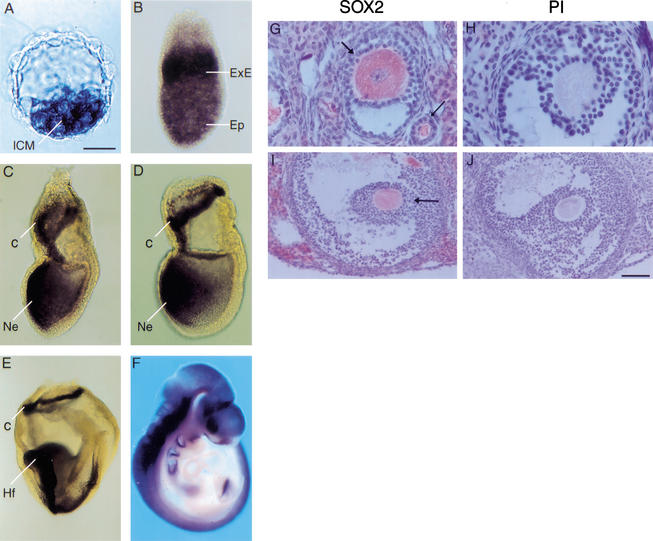
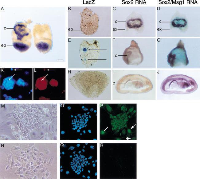
Each cell lineage specified in the preimplantation mammalian embryo depends on intrinsic factors for its development, but there is also mutual interdependence between them. OCT4 is required for the ICM/epiblast lineage, and at transient high levels for extraembryonic endoderm, but also indirectly through its role in regulating Fgf4 expression, for the establishment and proliferation of extraembryonic ectoderm from polar trophectoderm. The transcription factor SOX2 has also been implicated in the regulation of Fgf4 expression. We have used gene targeting to inactivate Sox2, examining the phenotypic consequences in mutant embryos and in chimeras in which the epiblast is rescued with wild-type ES cells. We find a cell-autonomous requirement for the gene in both epiblast and extraembryonic ectoderm, the multipotent precursors of all embryonic and trophoblast cell types, respectively. However, an earlier role within the ICM may be masked by the persistence of maternal protein, whereas the lack of SOX2 only becomes critical in the chorion after 7.5 days postcoitum. Our data suggest that maternal components could be involved in establishing early cell fate decisions and that a combinatorial code, requiring SOX2 and OCT4, specifies the first three lineages present at implantation.


Figures

Figure 1
Figure 1
Expression of Sox2 RNA in mouse embryos and SOX2 protein in oocytes. (AF) Whole-mount in situ hybridization in wild-type embryos. Sox2 RNA is seen in the ICM of the blastocyst (A), throughout the epiblast (Ep) and extraembryonic ectoderm (ExE) of a 6.5-dpc embryo (B), and in the chorion (c) and anterior region of the presumptive neuroectoderm (Ne) of a 7.5-dpc (C) and 7.5–8.0-dpc (D) embryo. (E) At 8.5 dpc, Sox2 expression persists in the chorion, headfolds (Hf), and neural tube. (F) By 9.5 dpc, it is seen throughout the nervous system, sensory placodes, branchial arches, and gut. Anterior is to the left in panels BE. (GJ) SOX2 antibody staining in wild-type adult ovary. Sections were incubated with SOX2 antiserum (G,I) or preimmune serum (PI; H,J). Staining is observed in the cytoplasm of oocytes in a developing primary follicle (G, thin arrow), an early antral follicle (G, thick arrow), and a mature antral follicle (I, arrow). Bars: A, 25 μm; BD, 60 μm; E, 120 μm; F, 435 μm; G,H, 20 μm; I,J, 53 μm.
Figure 2
Figure 2
Targeted disruption of Sox2. (A) Schematic diagram showing the Sox2 locus, β-geo targeting vector, and predicted fragment sizes of wild-type and disrupted clones. The restriction sites are EcoRI (RI), SalI (S), and NotI (N). The thick solid bar in red shows the vector homology regions. (B,C) Analysis of DNA from ES cell clones and offspring from Sox2βgeo heterozygous matings. DNA was digested with EcoRI, and a 3′ probe outside of the homology region was used on Southern blots (see A). TA, targeted allele. (DI) LacZ activity in Sox2βgeo heterozygous embryos. LacZ staining in a morula (2.5 dpc; D), blastocyst (3.5 dpc; E), 6.5-dpc embryo (F), 7.5-dpc embryo (G), 8.5-dpc embryo (H), and 9.5-dpc embryo (I). Bars: D,E, 25 μm; FH, 65 μm; I, 370 μm.
Figure 3
Figure 3
Sox2βgeo mutant embryos lack epiblast. (AH) Histological and marker analysis of 6.0-dpc embryos from Sox2βgeo intercrosses. (A,C,E,G) Normal embryos. (B,D,F,H) Mutant embryos. Sections were stained with haemotoxylin and eosin (H & E) in A and B. RNA in situ hybridization was performed for H19 (extraembryonic marker; C,D), Oct4 (epiblast marker; E,F), and Evx1 (VE marker; G,H). 35S-UTP-labeled probes were used for H19 and Oct4, and sections were counterstained with methyl green. A DIG-labeled probe was used for Evx1. The arrows shown in the main panel and inset of G refer to the same region. Evx1 staining in the wild-type embryo is distal to the embryonic/extraembryonic border as expected (G) and appears more punctate because the cells are flatter than those in the mutant embryo (H). Sox2βgeo null embryos form disorganized extraembryonic tissues, but do not form epiblast. Bars: A,B, 50 μm; CF, 65 μm; G (inset), 75 μm; G,H, 30 μm.
Figure 4
Figure 4
Sox2βgeo null blastocysts show defective ICM development in culture. (AH) Phase contrast microscopy of embryos from Sox2βgeo intercrosses, with wild-type (+/+; A) and Sox2βgeo null (−/−; E) blastocysts and different blastocyst outgrowths grown in culture for 3 d (BD,FH). Wild-type and mutant blastocysts look similar, but the ICM of null blastocysts fails to outgrow. (I) RT–PCR marker analysis of Sox2βgeo null blastocyst outgrowths. Morulae from Sox2βgeo heterozygous intercrosses were cultured for 5 d. BSC loading control refers to Bluescript RNA that was not added to an RNA preparation, but added directly to the subsequent RT reaction. No DNA refers to the negative control used during the PCR reaction. (JQ) Pl1 RNA in situ hybridization on Sox2βgeo null ICM outgrowths. Isolated ICMs (JM) were grown in culture for 4 d and analyzed for Pl1 expression (NQ). Mutant (P,Q) but not wild-type (N) or Sox2βgeo heterozygous (O) ICM outgrowths express PL1. Bars: A,E, 35 μm; BD,FH, 100 μm; JQ, 27 μm.
Figure 5
Figure 5
Nuclear and cytoplasmic staining of SOX2 protein in preimplantation embryos. (AL) SOX2 antibody staining in wild-type blastocysts. Embryos were incubated in SOX2 (B,H,L), preimmune (PI; E), or OCT4 (K) antiserum. Nuclei were stained with 7-AAD (A,D,G) or DAPI (J). (C,F,I) Overlays of the antibody and 7-AAD staining to show colocalization in yellow. (AF) Whole blastocysts. (GI) An ICM isolated by immunosurgery. (JL) Confocal images, using an affinity-purified SOX2 antibody. SOX2 is observed throughout the ICM in the nucleus (n) and cytoplasm (line) unlike OCT4, which is detected only in the nucleus. The arrowhead indicates the layer of ExEn where OCT4 is present but SOX2 levels are low. (MV) SOX2 antibody staining in wild-type embryos from 1-cell to late morula (L-morula) stages. Embryos were stained for SOX2 using affinity-purified antibody (MQ), and nuclei were visualized with DAPI (RV). All panels are confocal images. n, nuclear staining; c, cytoplasmic staining; pb, polar body. (W) SOX2 antibody staining in blastocysts from Sox2βgeo heterozygous intercrosses. Three separate litters were analyzed (13) using horseradish peroxidase and DAB staining. SOX2 is observed in all embryos. The last two embryos in each set are wild-type preimmune controls (*). Bars: AF,JL, 25 μm; GI, 30 μm; MV, 25 μm; W, 80 μm.
Figure 6
Figure 6
Chimera rescue of Sox2βgeo homozygotes and the presence of SOX2 protein in TS cells. (A) Sox2 RNA is absent in the chorion of a 7.5-dpc Sox2βgeo null homozygote rescued with wild-type ES cells (right) compared with a wild-type or heterozygote host embryo with chorionic (c) staining (left). (BJ) Chimeras were dissected to separate the embryo/epiblast (ep) from the extraembryonic region. The embryo was stained for lacZ (B,E,H), and extraembryonic tissue was first analyzed for Sox2 RNA by in situ hybridization (C,F,I) and then for Msg1 as a positive control (D,G,J). (BJ) Chimeras corresponding to two rescued Sox2βgeo homozygotes (E–J) and a wild-type host (based on Sox2 chorionic expression and no lacZ staining; BD). Arrows in E show Sox2βgeo null cells expressing lacZ in a rescued chimera. (K,L) SOX2 antibody staining in ES cells (L), and nuclear staining with DAPI (K). SOX2 is seen in the nucleus of ES cells (L, arrows), and is absent in STO feeder cells (K, large nuclei). (M,N) Phase micrographs of TS cells (M) and differentiated TS cells (N) grown in the absence of FGF4 for 4 d. (OR) SOX2 antibody staining in TS cells. SOX2 (P), preimmune (R), and DAPI (O,Q) staining. SOX2 is expressed in the nucleus of TS cells (round colony). In the surrounding differentiating cells, SOX2 remains nuclear (line), nuclear and cytoplasmic (arrow), or is greatly reduced (arrowhead), depending on the degree of differentiation. Bars: A, 125 μm; BJ, 90 μm; K,L, 23 μm; M,N, 15 μm; OR, 40 μm.

References

    1. Ambrosetti DC, Schöler HR, Dailey L, Basilico C. Modulation of the activity of multiple transcriptional activation domains by the HMG and POU domains mediates the synergistic action of Sox2 and Oct-3 on the FGF-4 enhancer. J Biol Chem. 2000;275:23387–23397. - PubMed
    1. Arman E, Haffner-Krausz R, Chen Y, Heath JK, Lonai P. Targeted disruption of fibroblast growth factor (FGF) receptor 2 suggests a role for FGF signaling in pregastrulation mammalian development. Proc Natl Acad Sci. 1998;95:5082–5087. - PMC - PubMed
    1. Avilion AA, Bell DM, Lovell-Badge R. Micro-capillary tube in situ hybridisation: A novel method for processing small individual samples. Genesis. 2000;27:76–80. - PubMed
    1. Beddington RSP, Robertson EJ. An assessment of the developmental potential of embryonic stem cells in the midgestation embryo. Development. 1989;105:733–737. - PubMed
    1. Botquin V, Hess H, Fuhrmann G, Anastassiadis C, Gross MK, Vriend G, Schöler HR. New POU dimer configuration mediates antagonistic control of an osteopontin preimplantation enhancer by Oct-4 and Sox-2. Genes & Dev. 1998;12:2073–2090. - PMC - PubMed

Publication types

MeSH terms