. 2003 Mar 14;278(11):9203-11.
doi: 10.1074/jbc.M209888200. Epub 2003 Jan 3.

Catalytic mechanism of thiol peroxidase from Escherichia coli. Sulfenic acid formation and overoxidation of essential CYS61

Affiliations

Catalytic mechanism of thiol peroxidase from Escherichia coli. Sulfenic acid formation and overoxidation of essential CYS61

Laura M S Baker et al. J Biol Chem. .

Abstract

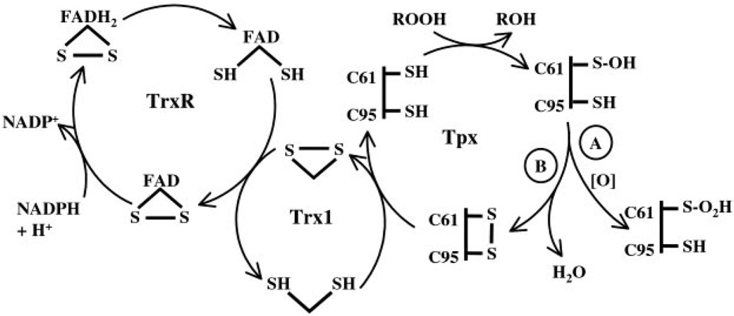
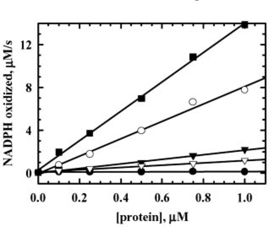
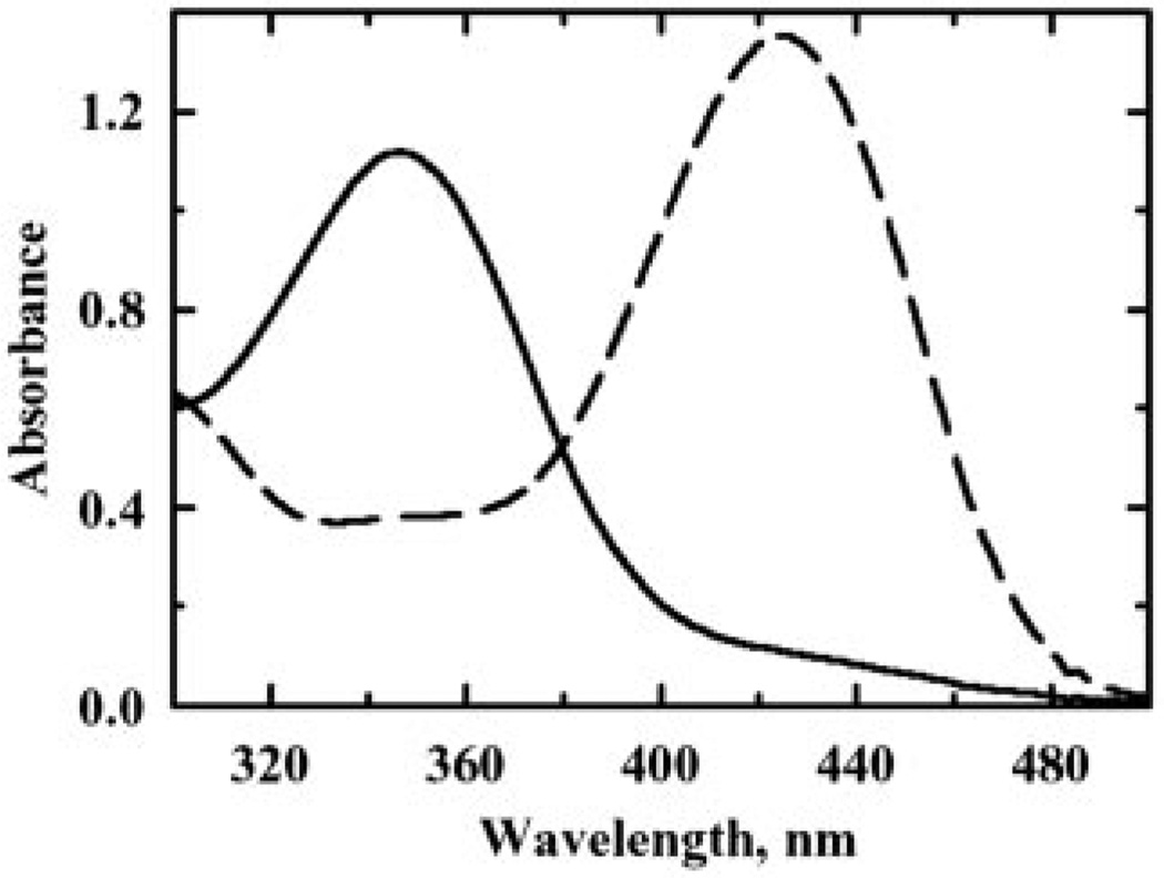
Escherichia coli thiol peroxidase (Tpx, p20, scavengase) is part of an oxidative stress defense system that uses reducing equivalents from thioredoxin (Trx1) and thioredoxin reductase to reduce alkyl hydroperoxides. Tpx contains three Cys residues, Cys(95), Cys(82), and Cys(61), and the latter residue aligns with the N-terminal active site Cys of other peroxidases in the peroxiredoxin family. To identify the catalytically important Cys, we have cloned and purified Tpx and four mutants (C61S, C82S, C95S, and C82S,C95S). In rapid reaction kinetic experiments measuring steady-state turnover, C61S is inactive, C95S retains partial activity, and the C82S mutation only slightly affects reaction rates. Furthermore, a sulfenic acid intermediate at Cys(61) generated by cumene hydroperoxide (CHP) treatment was detected in UV-visible spectra of 4-nitrobenzo-2-oxa-1,3-diazole-labeled C82S,C95S, confirming the identity of Cys(61) as the peroxidatic center. In stopped-flow kinetic studies, Tpx and Trx1 form a Michaelis complex during turnover with a catalytic efficiency of 3.0 x 10(6) m(-1) s(-1), and the low K(m) (9.0 microm) of Tpx for CHP demonstrates substrate specificity toward alkyl hydroperoxides over H(2)O(2) (K(m) > 1.7 mm). Rapid inactivation of Tpx due to Cys(61) overoxidation is observed during turnover with CHP and a lipid hydroperoxide, 15-hydroperoxyeicosatetraenoic acid, but not H(2)O(2). Unlike most other 2-Cys peroxiredoxins, which operate by an intersubunit disulfide mechanism, Tpx contains a redox-active intrasubunit disulfide bond yet is homodimeric in solution.


Figures

FIG. 1
FIG. 1. HPLC separation of tryptic peptides from either reduced or oxidized pyridylethylated Tpx and tryptic digest sites
Exhaustive digests of reduced (A and C) or oxidized (B and D) Tpx (5 nmol) that was pyridylethylated during denaturation prior to incubation with trypsin were fractionated on an AquaPore RP-300 C8 column. Gradient conditions are given under “Experimental Procedures,” and peptides were detected at 215 nm (A and B) and 254 nm (C and D). The closed arrows in each map indicate the following peaks: a pyridylethylated fragment containing Cys82 (74 min) (A and C) and disulfide-containing peaks P1 (68 min) and P2 (70 min) (B and D). The open arrows indicate fragments resulting from reduction of P1 or P2. E, a portion of the sequence of Tpx from E. coli (residues 45–113, accession number AAC74406) is shown with the tryptic sites (underlined) flanking the three Cys (boldface type) numbered from the initiating Met. The two Cys61 fragments resulting from the flanking tryptic sites are highlighted with different lines for P1 (dotted) and P2 (solid), and the total ESI-MS mass of each disulfide-containing peak (including the Cys95-containing fragment indicated by the dashed line) is given above the site of initial hydrolysis by trypsin.
FIG. 2
FIG. 2. Steady-state kinetic analysis of Tpx with various concentrations of CHP
All three protein components (TrxR (1.5 µm), Trx1 (10 µm), and Tpx (1 µm)) were incubated in 150 µm NADPH for 5 min in one syringe and then were mixed with varying amounts of CHP (0 µm (closed circles), 100 µm (open triangles), 200 µm (closed squares), and 400 µm (open diamonds)) in another syringe (final concentrations after mixing; see “Experimental Procedures”). Reaction progress was monitored at 340 nm on a stopped flow spectrophotometer at 25 °C, and rates were extrapolated from the linear portion of the curve (0–2 s) using linear regression analysis. At 100 µm CHP, Tpx was not inactivated and gave linear absorbance changes for the duration of the reaction and full peroxide consumption (open triangles). At higher CHP concentrations, Tpx activity diminished rapidly and nonlinearly without complete consumption of CHP or NADPH (closed squares and open diamonds). Stopped-flow data were collected every 50 ms, but only data from every 1.5 s are represented by the symbols.
FIG. 3
FIG. 3. Scheme of TrxR/Trx1 reduction and electron transfer pathways for Tpx during catalysis and inactivation
During Tpx turnover, a small proportion of Tpx becomes overoxidized and is removed from the reaction cycle (path A). The remaining Tpx reforms the redox-active disulfide to continue the catalytic cycle (path B). Eventually, after multiple turnovers in the presence of ≥150 µm CHP, the enzyme primarily converts to the R-SO2H species, few disulfide-containing species remain, and activity declines, leading to irreversible inactivation. This scheme depicts the overall flow of electrons but not necessarily the precise redox forms of TrxR involved in turnover.
FIG. 4
FIG. 4. Steady-state assay of Tpx as a function of 15-HPETE concentration
Reaction mixtures in one syringe containing NADPH (150 µm), Trx1 (10 µm), TrxR (1.5 µm), and Tpx (1 µm) were mixed with varying amounts of 15-HPETE (0–30 µm) in another syringe on the stopped-flow spectrophotometer at 25 °C (final concentrations after mixing). Each rate is the average of three experiments and was obtained by linear regression analysis of the linear portion of the reaction progression (0–1 s) prior to inactivation. Standard peroxidase buffer was used in all cases as described under “Experimental Procedures.”
FIG. 5
FIG. 5. Steady-state kinetic analysis of wild type and mutant Tpx proteins
Tpx reaction mixtures in one syringe containing NADPH (150 µm), Trx1 (10 µm), TrxR (1.5 µm), and varying amounts (0–1 µm) of Tpx (closed squares), C82S (open circles), C95S (closed triangles), C82S,C95S (open triangles), or C61S (closed circles) were mixed with CHP (50 µm) on the stopped-flow spectrophotometer in standard peroxidase buffer (final concentrations after mixing; see “Experimental Procedures”). Each rate is the average of three experiments conducted at 25 °C and was obtained by linear regression analysis of the linear portion of the reaction (0–1 s) prior to inactivation.
FIG. 6
FIG. 6. Spectrophotometric analysis of NBD-labeled Tpx mutants
Prereduced C82S,C95S treated with 1 eq of cumene hydroperoxide (solid line) or no peroxide (dashed line) was modified with NBD chloride (10×) for 5 min. To remove excess reagent, treated samples were washed with 5 ml of buffer by ultrafiltration, and then labeled proteins were analyzed from 200 to 600 nm.

References

    1. Storz G, Tartaglia LA, Farr SB, Ames BN. Trends Genet. 1990;6:363–368. - PubMed
    1. Carmel-Harel O, Storz G. Annu. Rev. Microbiol. 2000;54:439–461. - PubMed
    1. Halliwell B, Gutteridge JMC. Free Radicals in Biology and Medicine. 3rd Ed. Oxford: Oxford University Press; 1999. pp. 188–276.
    1. Storz G, Imlay JA. Curr. Opin. Microbiol. 1999;2:188–194. - PubMed
    1. Lynch AS, Lin ECC. In: E. coli and Salmonella: Cellular and Molecular Biology VI. 2nd Ed. Neidhardt FC, editor. Washington, D. C.: American Society for Microbiology Press; 1996. pp. 1526–1538.

Publication types

MeSH terms

LinkOut - more resources