. 2003 Jan 1;23(1):1-6.
doi: 10.1523/JNEUROSCI.23-01-00002.2003.

Actin-ATP hydrolysis is a major energy drain for neurons

Affiliations

Actin-ATP hydrolysis is a major energy drain for neurons

Barbara W Bernstein et al. J Neurosci. .

Abstract

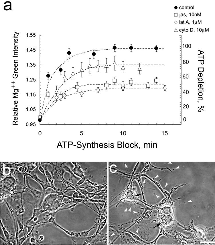
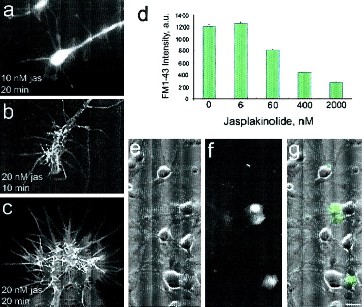
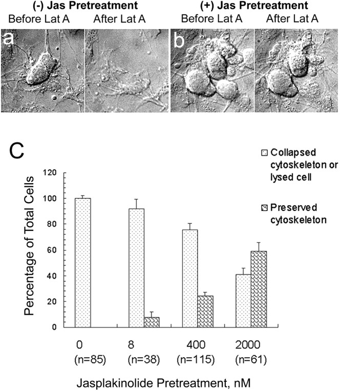
In cultured chick ciliary neurons, when ATP synthesis is inhibited, ATP depletion is reduced approximately 50% by slowing actin filament turnover with jasplakinolide or latrunculin A. Jasplakinolide inhibits actin disassembly, and latrunculin A prevents actin assembly by sequestering actin monomers. Cytochalasin D, which allows assembly-disassembly, but only at pointed ends, is less effective in conserving ATP. Ouabain, an Na(+)-K(+)-ATPase inhibitor, and jasplakinolide both prevent approximately 50% of the ATP loss. When applied together, they completely prevent ATP loss over a period of 20 min, suggesting that filament stabilization reduces ATP consumption by decreasing actin-ATP hydrolysis directly rather than indirectly by modulating the activity of Na(+)-K(+)-ATPase, a major energy consumer.


Figures

Fig. 1.
Fig. 1.
a, Slowing filament turnover conserves ATP. Time-lapse imaging of ciliary neurons, loaded with MgGr, was used to monitor ATP depletion after a block in ATP synthesis. As ATP is depleted, [Mg2+]i increases. Analysis includes all cells in 20 fields. Similar results were seen in two replicas of this experiment; mean ± SEM of >100 cells.b, c, Phase micrographs of fixed neurons before (b) and 40 min after (c) application of ATP-synthesis block (10 mm NaN3 to 6 mm 2-deoxyglucose). Chemically ischemic cells show extensive neurite retraction, somata shrinking, and rounding, lysed cell debris (arrowheads), and vacuoles (arrows). Scale bar, 30 μm.jas, Jasplakinolide; lat A, latrunculin A; cyto D, cytochalasin D.
Fig. 2.
Fig. 2.
The minimum jasplakinolide concentration needed to stabilize filaments is >6 nm. a–c, Incubation in as little as 20 nm jasplakinolide (jas) induces a filopodial expansion, indicating stimulation of actin assembly rather than mere stabilization of F-actin, here stained with rhodamine phalloidin. Scale bar, 10 μm.d, Transmitter vesicle recycling depends on filament turnover rate (Bernstein et al., 1998) and is attenuated by preincubation in jasplakinolide >6 nm. Cells were depolarized in a 75 mm K+ buffer containing the fluorescent styryl dye FM1-43 (10 μm), which is used to monitor depolarization-induced vesicle cycling (Cochilla et al., 1999). g, Overlay of DIC (e) and fluorescence (f) images, showing depolarization-induced FM1-43 uptake in ciliary calyx. Scale bar, 20 μm.
Fig. 3.
Fig. 3.
Morphological evidence for promotion of actin disassembly by latrunculin (Lat A) and stabilization of F-actin by jasplakinolide (Jas). a, DIC micrographs of live cells without jasplakinolide pretreatment show lysis and loss of cell shape (flattening) after 15 min of incubation in 1 μm latrunculin A. The dose-dependent nature of jasplakinolide stabilization of cell morphology seen inb is plotted in c. A concentration as low as 8 nm has a stabilizing effect. All cells in >20 fields were included in data plotted for each jasplakinolide concentration.
Fig. 4.
Fig. 4.
Effects of actin modulators on ATP, Na+, and Ca2+ levels in ischemically stressed cells. a, Reducing filament turnover moderates ischemically induced increase in [Ca2+]i but is not likely to contribute significantly to ATP conservation of jasplakinolide (jas)- and latrunculin A (lat A)-treated cells. b, Similar time courses were observed for ATP depletion in rat embryonic day 18 brain cortical cells measured with either a cell lysate–luciferase assay or an [Mg2+]i fluorescent dye indicator and for ciliary cells using MgGr or Mag-fura-2; exponential curve fit shown. c, d, Actin assembly modulators conserve ATP via a reduction in actin-ATP hydrolysis rather than a reduction in Na+–K+-ATPase activity.c, Preincubation in 10 nm jasplakinolide or 1 μm latrunculin A accelerates [Na+]i increase during ATP depletion. Only in ATP-depleted control cells is [ATP] low enough to reverse Na+–Ca2+ exchanger, pulling Ca2+ in and extruding Na+. Not unexpectedly, PBS–1.5 mm ouabain, an Na+–K+-ATPase inhibitor, causes the fastest rise in Na+. d, Conservation of ATP by ouabain and jasplakinolide is additive.a–d, Mean ± SEM of two or three experiments;n = 200–300 cells in 60 fields.

References

    1. Alberts B, Bray D, Raff M, Roberts K, Watson JD. Molecular biology of the cell, pp 41–88. Garland; London: 1994.
    1. Aliev G, Smith MA, Seyidov D, Neal ML, Lamb BT, Nunomura A, Gasimov EK, Vinters HV, Perry G, LaManna JC, Friedland RP. The role of oxidative stress in the pathophysiology of cerebrovascular lesions in Alzheimer's disease. Brian Pathol. 2002;12:21–35. - PMC - PubMed
    1. Attwell D, Laughlin SB. An energy budget for signaling in the grey matter of the brain. J Cereb Blood Flow Metab. 2001;21:1133–1145. - PubMed
    1. Bamburg JR. Proteins of the ADF/cofilin family: essential regulators of actin dynamics. Annu Rev Cell Dev Biol. 1999;15:185–230. - PubMed
    1. Belmont LD, Drubin DG. The yeast V159N actin mutant reveals roles for actin dynamics in vivo. J Cell Biol. 1998;142:1289–1299. - PMC - PubMed

Publication types

MeSH terms

LinkOut - more resources