. 2003 Jan 1;23(1):131-40.
doi: 10.1523/JNEUROSCI.23-01-00131.2003.

KIF17 dynamics and regulation of NR2B trafficking in hippocampal neurons

Affiliations

KIF17 dynamics and regulation of NR2B trafficking in hippocampal neurons

Laurent Guillaud et al. J Neurosci. .

Abstract

KIF17, a recently characterized member of the kinesin superfamily proteins, has been proposed to bind in vitro to a protein complex containing mLin10 (Mint1/X11) and the NR2B subunit of the NMDA receptors (NMDARs). In the mammalian brain, NMDARs play an important role in synaptic plasticity, learning, and memory. Here we present, for the first time, the dynamic properties of KIF17 and provide evidence of its function in the transport of NR2B in living mammalian neurons. KIF17 vesicles enter and move specifically along dendrites in a processive way, at an average speed of 0.76 microm/sec. These vesicles are effectively associated with extrasynaptic NR2B, and thus they transport and deliver NR2B subunits in dendrites. However, KIF17 does not seem to enter directly into postsynaptic regions. Cellular knockdown or functional blockade of KIF17 significantly impairs NR2B expression and its synaptic localization. Interestingly, the decrease in the number of synaptic NR2B subunits is followed by a parallel increase in the number of NR2A subunits at synapses. In contrast, upregulation of the expression level of NR2B, after treatment with the NMDAR antagonist D(-)-2-amino-5-phosphonopentanoic acid, simultaneously increases the expression level of KIF17. These observations concerning the downregulation or upregulation of KIF17 and NR2B reveal the probable existence of a shared regulation process between the motor and its cargo. Taken together, these results illustrate the complex mechanisms underlying the active transport and regulation of NR2B by the molecular motor KIF17 in living hippocampal neurons.


Figures
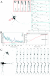
Fig. 1.
Fig. 1.
Dynamic properties of YFP-KIF17 in living hippocampal neurons. Hippocampal neurons cultured for 10 d were transfected with YFP-KIF17. After 24 hr, the living cells were observed under a confocal laser-scanning microscope. All images were inverted to improve visibility. A, Fluorescence imaging of FRAP and movement of YFP-KIF17 in transfected neurons. A 15 μm area on the apical dendrite (red inset) was bleached, and the fluorescence recovery was monitored every 5 sec. Recovery was achieved from both the proximal part (P) and the distal part (D) of the bleached area. Small vesicular structures moved within the area, and fluorescence was completely restored after 20 sec. Movement of individual YFP-KIF17 vesicles was also monitored over time in another dendrite of the same neuron (blue inset). Images were taken every 3 sec and show the movement of two vesicles (*) from the proximal (P) to the distal (D) part of the dendrite. Initial positions 1 and2 and final positions 1′ and2′ of both vesicles were plotted below. Scale bar, 20 μm. B, Graphic analysis of FRAP experiments. Shown is a plot profile of the fluorescence intensity as a function of the distance in the bleached area (left panel). An increasing number of fluorescence peaks corresponding to vesicular structures of YFP-KIF17 appeared in the bleached area. Mean fluorescence intensity as a function of time plotted on theright panel shows the initial fluorescence intensity (Ii), the fluorescence intensity right after bleaching (I0), the final fluorescence intensity (If) after complete recovery, and the half fluorescence intensity (I1/2). PB, Prebleach; B, bleach; R, recovery.C, High frame-rate acquisition of a transfected hippocampal neuron expressing YFP-KIF17. Several vesicles moved anterogradely, whereas few aggregates were immobile. D, High frame-rate acquisition of a transfected hippocampal neuron expressing YFP-KIF17 and treated with 20 μm nocodazole for 45 min. The movement of YFP-KIF17 is inhibited completely. Increasing numbers of immobile aggregates can be observed both in dendrites and in the cell body.
Fig. 2.
Fig. 2.
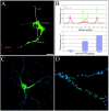
Distribution of YFP-KIF17 in hippocampal neurons.A, Dendritic localization of YFP-KIF17. Transfected hippocampal neurons were immunostained with anti-phosphorylated-NF-H mAb to discriminate axon (red) from dendrites. YFP-KIF17 (green) localizes mainly in dendrites and the cell body. Scale bar, 20 μm. B, Quantitative analysis of the axo/dendritic distribution of YFP-KIF17. The fluorescence intensity profile showing YFP-KIF17 (green) and NF-H (red) was measured using the fluorescence profile function of LSM510 software on an x–z plane following the line to obtain a profile containing cross sections of the axon, the cell body, and the dendrite at the same time.C, D, Three-dimensional reconstruction images of hippocampal neurons expressing YFP-KIF17. Ten-day-old cultures were transfected with YFP-KIF17 (green) and processed for immunodetection of synaptic clusters using anti-PSD95 mAb (blue). Rendering of 3D images from confocal Z stacks files was performed using Autodeblur and Autovisualize. YFP-KIF17 is fully restricted to the dendritic shaft and did not enter in postsynaptic regions.
Fig. 3.
Fig. 3.
Distribution of YFP-mLin10 in HA-KIF17-cotransfected hippocampal neurons. After 10 d of culture, hippocampal neurons were cotransfected with HA-KIF17 and YFP-mLin10. Twenty-four hours after transfection, the cells were fixed (see Materials and Methods), and HA-KIF17 was detected with the Alexa568 fluorescent secondary antibody. Overexpression of HA-KIF17 (red) induced a clear redistribution of YFP-mLin10 (green) from its initial perinuclear localization (as observed in control single-transfected cells) to the dendritic network. Scale bar, 20 μm. Statistical analysis of the redistribution of YFP-mLin10 in hippocampal neurons shows the percentage of cells with perinuclear or dendritic localization of YFP-mLin10 in single-transfected cells or HA-KIF17 cotransfected cells.Undefined corresponds to an unclear pattern with both perinuclear and dendritic localizations of the proteins.
Fig. 4.
Fig. 4.
Colocalization of YFP-KIF17 and NR2B. After 10 d of culture, hippocampal neurons were transfected with YFP-KIF17, and living cells were observed under a confocal laser-scanning microscope. Vesicular movement of YFP-KIF17 was first verified in the transfected neurons. The cells were then fixed, and immunodetection was performed as described in Materials and Methods.A, Transfected neuron showing YFP-KIF17 (green) and NR2B mAb (red). Shown is colocalization (yellow) of YFP-KIF17 and NR2B on the same vesicles (arrowheads). Higher magnification is shown in the blue inset. Scale bar, 20 μm.B, C, Localization of extrasynaptic and synaptic NR2B clusters. Hippocampal neurons expressing YFP-KIF17 (green) were permeabilized and immunostained (see Materials and Methods) for PSD95 mAb (blue) and NR2B pAb (red) (B) or synaptophysin mAb (blue) and NR2B pAb (red) (C). The mobile extrasynaptic NR2B subunits colocalize with YFP-KIF17 vesicles (yellow andarrowheads), whereas the immobile synaptic NR2B subunits colocalize with PSD95 or synaptophysin (pink). No colocalization of YFP-KIF17, PSD95, and NR2B or synaptophysin and NR2B was observed.
Fig. 5.
Fig. 5.
Inhibition of KIF17 in hippocampal neurons.A, Inhibition of KIF17 and NR2B expression by antisense oligonucleotide treatment. Hippocampal neurons were exposed (+) to 1 μm antisense or sense oligonucleotides against KIF17 for 3 d. Oligonucleotides were then washed out (−), and neurons were further cultured for 3 more days. After the cells were harvested, 20 μg of protein was separated by PAGE and analyzed by Western blotting with antibodies against KIF17, NR2B, and KIF5B. Bands were detected using ECL, films were scanned, and the bands were quantified using ImageJ. All measurements were standardized on tubulin content. Antisense oligonucleotide treatment (green curves) completely inhibited KIF17 expression and also led to a net decrease in the NR2B expression level. No inhibition was observed after sense oligonucleotide treatment (red curves). KIF5B expression was not affected by antisense or sense oligonucleotide treatment. After recovery from antisense oligonucleotide treatment, both KIF17 and NR2B expressions were restored. Insets on each plot show the bands detected by Western blot analysis (*) after treatment with sense (S) or antisense (A) oligonucleotides for KIF17, NR2B, and KIF5B. B, Western blot analysis of related proteins was performed as described above. Proteins were detected with specific antibodies (see Materials and Methods). Antisense oligonucleotide treatment completely inhibited KIF17 expression and reduced NR2B and mLin10 expression levels by 33 and 64%, respectively. NR2A expression level increased by 24%. No changes in expression level were observed for tubulin, KIF1A, NR2C, PSD95, and GluR1. C, Distribution of NR2B in KIF17-knocked-down hippocampal neurons. After 10 d of culture, hippocampal neurons were exposed to 1 μmFITC-tagged antisense oligonucleotides against KIF17. Twenty-four hours later, the cells were fixed, permeabilized, and processed for immunolocalization of NR2B as described in Materials and Methods. NR2B clusters were counted in a 20 μm area. Hippocampal neurons transfected with FITC-tagged antisense oligonucleotides (green) showed a marked decrease in the synaptic distribution of NR2B clusters (red). Higher magnification of control neurons or neurons exposed to antisense oligonucleotides (FITC-AS) is shown. Scale bar, 20 μm.D, Distribution of NR2B in dominant-negative overexpressing hippocampal neurons. After 10 d of culture, hippocampal neurons were transfected with YFP-610. Twenty-four hours later, the cells were fixed and processed for immunolocalization of NR2B. Hippocampal neurons transfected with YFP-610 (green) showed a similar decrease in the synaptic distribution of NR2B clusters (red), as observed after FITC-AS treatment. Note that accumulation of NR2B can be observed in the cell body of YFP-610-transfected neurons. Higher magnification of control neurons or YFP-610-overexpressing neurons is shown. Scale bar, 20 μm.
Fig. 6.
Fig. 6.
Distribution of NR2 subunits in hippocampal neurons. After 10 d of culture, hippocampal neurons were transfected with YFP-610, fixed, and processed for immunolocalization of NR2A, NR2B, NR2C, and PSD95. The number of positive clusters for each subunit and for PSD95 was counted in a 20 μm area. A 23% decrease in the number of NR2B clusters and an 18% increase in the number of NR2A clusters were observed in neurons overexpressing YFP-610 compared with control nontransfected neurons. No significant changes were observed for NR2C. The total number of PSD95 synaptic clusters did not change.
Fig. 7.
Fig. 7.
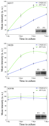
Upregulation of KIF17 and NR2B in neuronal culture. Hippocampal neurons were stimulated with either 100 μm AP-V or 10 μm CNQX up to 7 d of culture. After the cells were harvested, 20 μg of proteins was separated by PAGE and analyzed by Western blotting with antibodies against KIF17, NR2B, and KIF5B. Proteins were detected by ECL, films were scanned, and the bands were quantified using ImageJ. All measurements were standardized on tubulin content. After stimulation with AP-V (green curves), a twofold increase in NR2B and KIF17 expression levels was observed, whereas no change in the expression level of KIF5B was observed. KIF17, NR2B, and KIF5B expression levels did not change after CNQX simulation (blue curves).Insets on each plot show the bands detected by Western blot analysis after 5 d (*) treatment with AP-V (A) or CNQX (C) for KIF17, NR2B, and KIF5B.

References

    1. Baas PW, Deitch JS, Black MM, Banker GA. Polarity orientation of microtubules in hippocampal neurons: uniformity in the axon and nonuniformity in the dendrite. Proc Natl Acad Sci USA. 1988;85:8335–8339. - PMC - PubMed
    1. Barria A, Malinow R. Subunit-specific NMDA receptor trafficking to synapses. Neuron. 2002;35:345–353. - PubMed
    1. Bliss TV, Collingridge GL. A synaptic model of memory: long-term potentiation in the hippocampus. Nature. 1993;361:31–39. - PubMed
    1. Burack MA, Silverman MA, Banker G. The role of selective transport in neuronal protein sorting. Neuron. 2000;26:465–472. - PubMed
    1. Craven SE, El-Husseini AE, Bredt DS. Synaptic targeting of the postsynaptic density protein PSD-95 mediated by lipid and protein motifs. Neuron. 1999;22:497–509. - PubMed

Publication types

MeSH terms

LinkOut - more resources