. 2003 Jan 1;23(1):230-9.
doi: 10.1523/JNEUROSCI.23-01-00230.2003.

Normal female sexual development requires neuregulin-erbB receptor signaling in hypothalamic astrocytes

Affiliations

Normal female sexual development requires neuregulin-erbB receptor signaling in hypothalamic astrocytes

Vincent Prevot et al. J Neurosci. .

Abstract

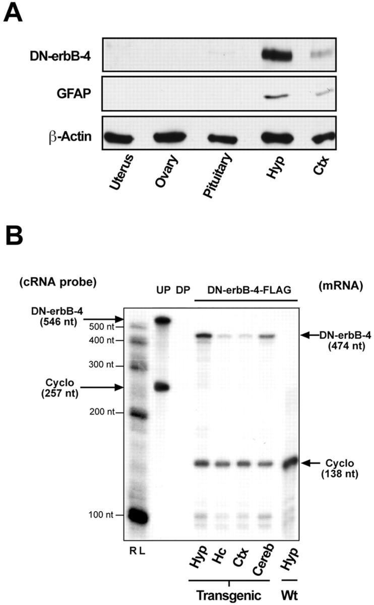
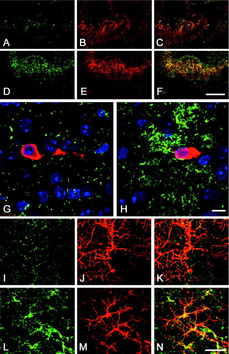
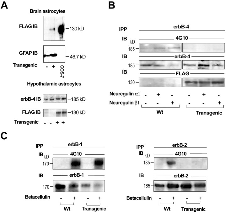
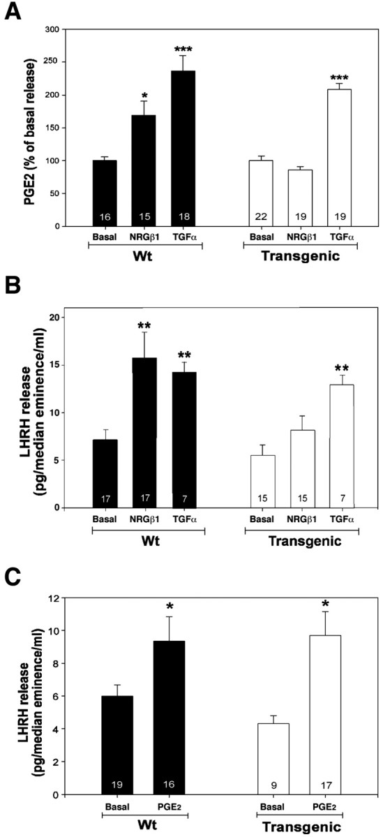
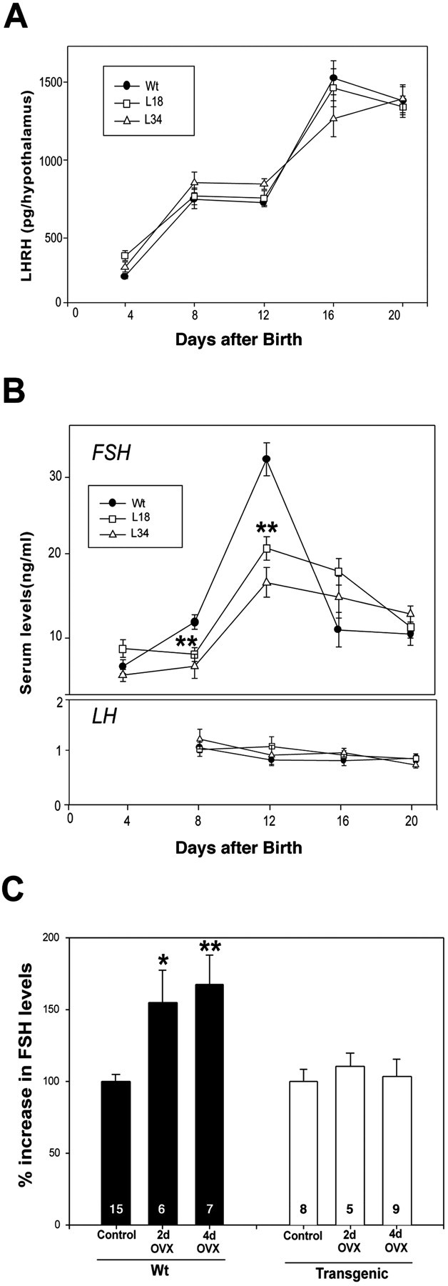
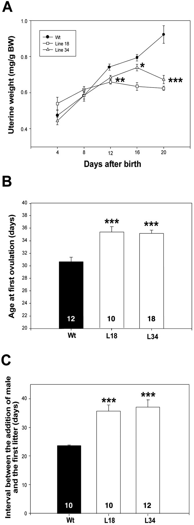
The initiation of mammalian puberty requires the activation of hypothalamic neurons secreting the neuropeptide luteinizing hormone-releasing hormone (LHRH). It is thought that this activation is caused by changes in trans-synaptic input to LHRH neurons. More recently, it has been postulated that the pubertal increase in LHRH secretion in female animals also requires neuron-glia signaling mediated by growth factors of the epidermal growth factor (EGF) family and their astrocytic erbB receptors. Although it appears clear that functional astrocytic erbB1 receptors are necessary for the timely advent of puberty, the physiological contribution that erbB4 receptors may make to this process has not been established. To address this issue, we generated transgenic mice expressing a dominant-negative erbB4 receptor (DN-erbB4) under the control of the GFAP promoter, which targets transgene expression to astrocytes. DN-erbB4 expression is most abundant in hypothalamic astrocytes, where it blocks the ligand-dependent activation of glial erbB4 and erbB2 receptors, without affecting erbB1 (EGF) receptor signaling. Mice carrying the transgene exhibit delayed sexual maturation and a diminished reproductive capacity in early adulthood. These abnormalities are related to a deficiency in pituitary gonadotropin hormone secretion, caused by impaired release of LHRH, the hypothalamic neuropeptide that controls sexual development. In turn, the reduction in LHRH release is caused by the inability of hypothalamic astrocytes to respond to neuregulin (NRG) with production of prostaglandin E(2), which in wild-type animals mediates the stimulatory effect of astroglial erbB receptor activation on neuronal LHRH release. Thus, neuron-astroglia communication via NRG-erbB4/2 receptor signaling appears to be essential for the timely unfolding of the developmental program by which the brain controls mammalian sexual maturation.


Figures

Fig. 1.
Fig. 1.
Analysis of transgene expression by Western blot and RNase protection assays. A, DN–erbB4 protein is expressed in both hypothalamic and cortical tissues but not in peripheral organs involved in reproductive function. Notice that the relative abundance of both DN–erbB4 and GFAP proteins is much greater in the hypothalamus than in the cerebral cortex. β-Actin was used as a loading control. Hyp, Hypothalamus;Ctx, cerebral cortex. B, The content of DN–erbB4 mRNA detected by RNase protection assay is greater in the hypothalamus than in other brain regions. RL, RNA ladder; UP, undigested probe; DP, digested probe; Cyclo, cyclophilin; Hc, hippocampus; Cereb, cerebellum; Wt, wild type.
Fig. 2.
Fig. 2.
Analysis of DN–erbB4 expression in the hypothalamus of 12-d-old mice by confocal microscopy.A–F, erbB4 immunoreactivity in astrocytes of the median eminence–medial basal hypothalamus of wild-type mice (A–C) and DN–erbB4 transgenic mice (D–F). A, D, erbB4 staining (green). B, E, GFAP staining (red). C, F, Merged images of erbB4–GFAP immunoreactive cells. G, H, Astrocytes of the preoptic area, which contains the cell bodies of LHRH neurons (red), show a much greater abundance of immunoreactive erbB4 (green) in DN–erbB4 mutant mice (H) than in wild-type animals (G). I–N, In DN–erbB4 mice, erbB4 immunoreactivity (green) is more abundant in hypothalamic astrocytes (L–N) than in astrocytes of the cerebral cortex (I–K). Astrocytes are identified by their GFAP immunoreactivity (red; J, M). Merged images for each brain area are shown in K (cerebral cortex) andN (hypothalamus). Scale bars: A–F, 100 μm; G, H, 10 μm; I–N, 5 μm.
Fig. 3.
Fig. 3.
DN–erbB4 expression in astrocytes blocks ligand-induced erbB4 and erbB2 receptor phosphorylation without affecting erbB1-mediated signaling. A, DN–erbB4 is expressed in cultured astrocytes isolated from either the whole brain (top panel) or the hypothalamus (bottom panel). Cell homogenates prepared from primary astrocyte cultures or COS-7 cells transfected with DN–erbB4 FLAG were subjected to SDS-PAGE and then immunoblotted (IB) with FLAG, erbB4, or GFAP antibodies.B, Inhibition of erbB4 receptor phosphorylation by the transgenic expression of DN–erbB4 in whole brain astrocyte cultures. Cells were treated with NRGβ1 (50 ng/ml for 5 min), lysed, immunoprecipitated (IPP) with erbB4 antibodies, subjected to SDS-PAGE separation, and blotted with 4G10 anti-phosphotyrosine antibodies (top panel). The membrane was stripped and incubated with an antibody that recognizes the intracellular domain of erbB4, showing the relative amount of receptor immunoprecipitated in each sample (middle panel). After stripping again, the membrane was incubated with an anti-FLAG antibody, showing that DN–erbB4 was only expressed in mutant cells, and that the level of expression was similar in all conditions. C, DN–erbB4 expression does not affect ligand-induced erbB1 receptor phosphorylation but abolishes erbB2 phosphorylation. Brain astrocyte cultures were treated with betacellulin (50 ng/ml for 5 min), lysed, and immunoprecipitated with either erbB1 (left panel) or erbB2 (right panel) antibodies, before SDS-PAGE and immunoblotting with 4G10 antiphosphotyrosine antibodies, followed by stripping and immunoblotting with either erbB1 or erbB2 antibodies to ensure that each receptor had been immunoprecipitated. Wt, Wild type.
Fig. 4.
Fig. 4.
DN–erbB4 expression suppresses NRG-induced release of LHRH from median eminence nerve terminals and PGE2 from hypothalamic astrocytes. A, NRGβ1-induced PGE2 release from primary cultures of hypothalamic astrocytes is abolished in DN–erbB4 mutants, whereas TGFα-stimulated PGE2 release is not altered [basal PGE2 release was similar in wild-type and DN–erbB4 cultures (238 ± 13 vs 223 ± 16 pg/ml, respectively)].B, DN–erbB4 expression suppresses NRGβ1-induced but not TGFα-induced LHRH release from the median eminence. C, PGE2 treatment (1 μm for 30 min) stimulates LHRH release from the median eminence of both wild-type and transgenic mice incubated in Krebs–Ringer bicarbonate buffer. In all panels, *p < 0.05, **p < 0.01, ***p < 0.001 versus basal release.Bars are means, vertical lines are the SEM, and the numbers inside the bars are the number of independent observations per group. Wt, Wild type.
Fig. 5.
Fig. 5.
Transgenic mice exhibit a normal content of LHRH in the hypothalamus but have reduced serum FSH levels and a reduced FSH secretory response to removal of ovarian steroid inhibitory control during the infantile period of development. A, Hypothalamic LHRH content measured by radioimmunoassay is similar in wild-type and mutant mice throughout neonatal–infantile development.B, The infantile increase in serum FSH levels that characterizes early postnatal development of the hypothalamic–pituitary unit in rodents is blunted in transgenic mice (top panel), whereas the low serum LH levels observed at this time remain unchanged (bottom panel) (10–20 mice per point). C, Ovariectomy (OVX) on postnatal day 12 results in a significant increase in serum FSH levels 2 and 4 d later in wild-type mice but not in mutant mice. Numbers inside the bars are the number of mice per group. Vertical lines are the SEM. In B, **p < 0.01 versus the wild-type group. InC, *p < 0.05 and **p < 0.01 between ovariectomized and nonovariectomized wild-type groups. Wt, Wild type.
Fig. 6.
Fig. 6.
Transgenic mice have retarded uterine growth, delayed sexual development, and reduced reproductive capacity in early adulthood. A, Uterine growth is reduced in mutant mice (8–12 animals per point, except day 12, in which each point has 30–44 mice). B, The age at first ovulation is delayed in transgenic mice. C, The age at first pregnancy is delayed in transgenic mice. In all panels, *p < 0.05, **p < 0.01, and ***p < 0.001 versus wild-type values.Circles, triangles, orbars are means; vertical lines represent the SEM. L18 and L34 are transgenic lines. Wt, Wild-type mice. Numbers inside the bars are the number of animals per group.

References

    1. Besnard F, Brenner M, Nakatani Y, Chao R, Purohit HJ, Freese E. Multiple interacting sites regulate astrocyte-specific transcription of the human gene for glial fibrillary acidic protein. J Biol Chem. 1991;266:18877–18883. - PubMed
    1. Brenner M, Kisseberth WC, Su Y, Besnard F, Messing A. GFAP promoter directs astrocyte-specific expression in transgenic mice. J Neurosci. 1994;14:1030–1037. - PMC - PubMed
    1. Campbell WB, Ojeda SR. Measurement of prostaglandins by radioimmunoassay. Methods Enzymol. 1987;141:323–350. - PubMed
    1. Chen MS, Bermingham-McDonogh O, Danehy FT, Jr, Nolan C, Scherer SS. Expression of multiple neuregulin transcripts in postnatal rat brains. J Comp Neurol. 1994;349:389–400. - PubMed
    1. Corfas G, Rosen KM, Aratake H, Krauss R, Fischbach GD. Differential expression of ARIA isoforms in the rat brain. Neuron. 1995;14:103–115. - PubMed

Publication types

MeSH terms