. 2003 Jan 1;23(1):260-8.
doi: 10.1523/JNEUROSCI.23-01-00260.2003.

Signaling by bone morphogenetic proteins and Smad1 modulates the postnatal differentiation of cerebellar cells

Affiliations

Signaling by bone morphogenetic proteins and Smad1 modulates the postnatal differentiation of cerebellar cells

Catherine Angley et al. J Neurosci. .

Abstract

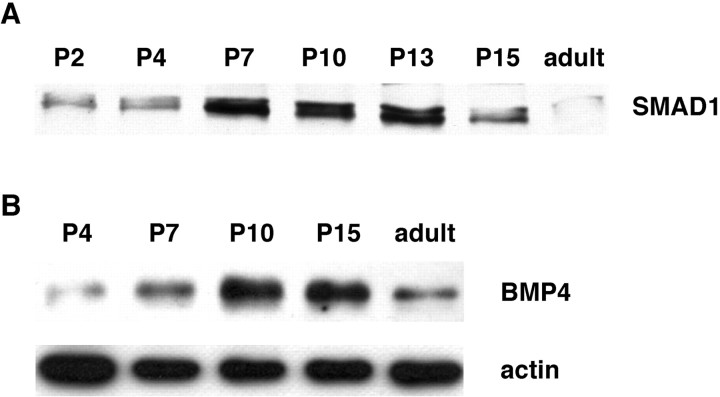
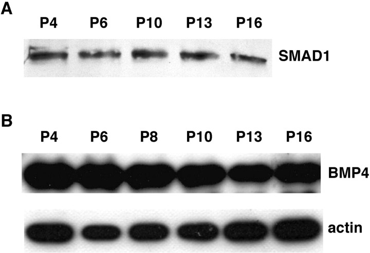
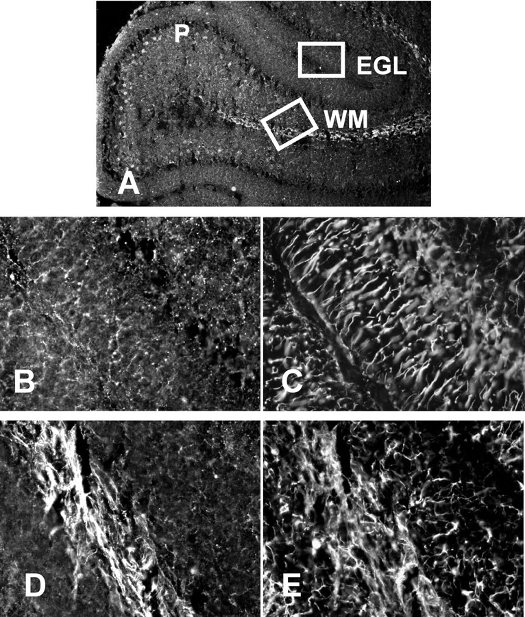
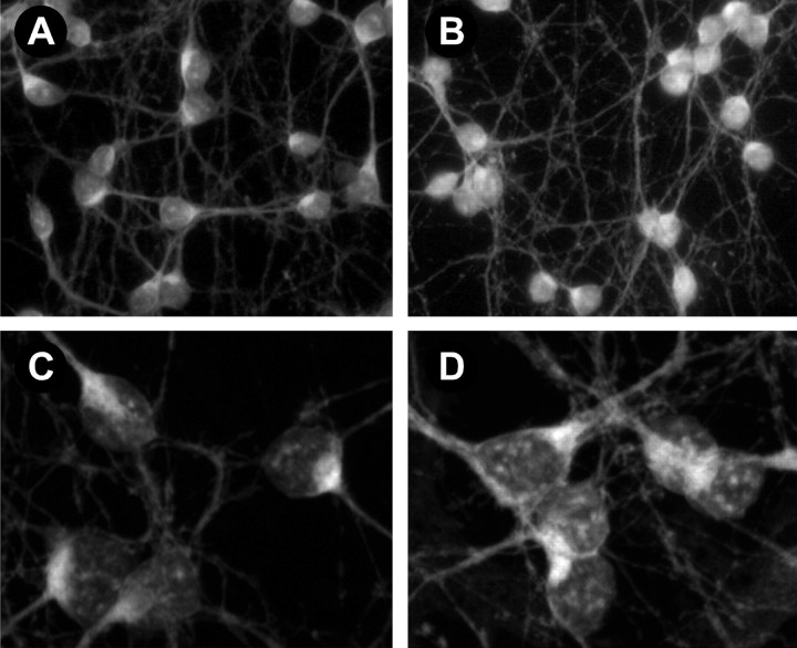
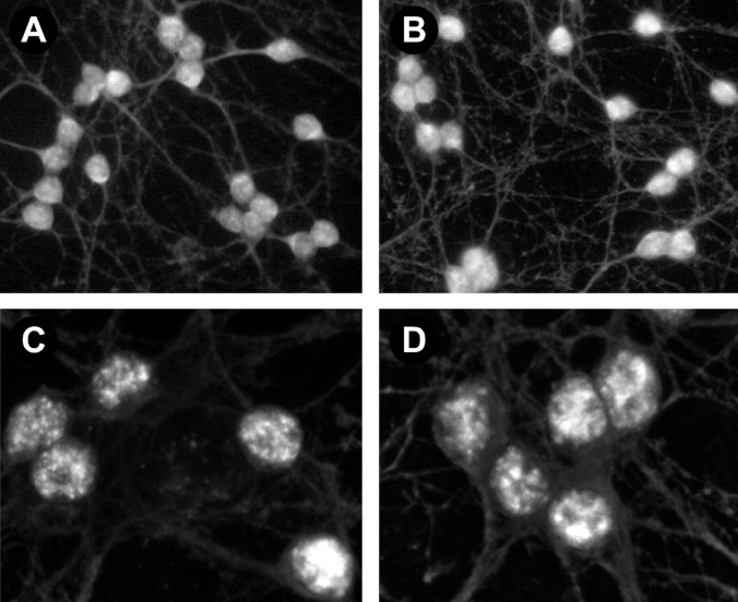
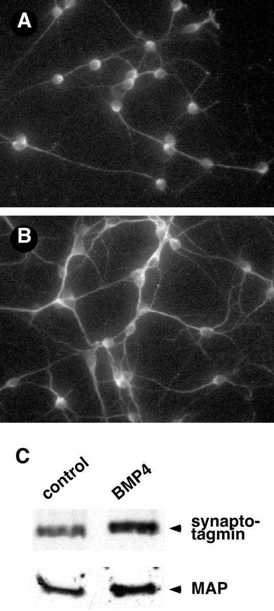
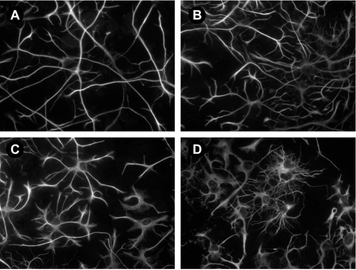
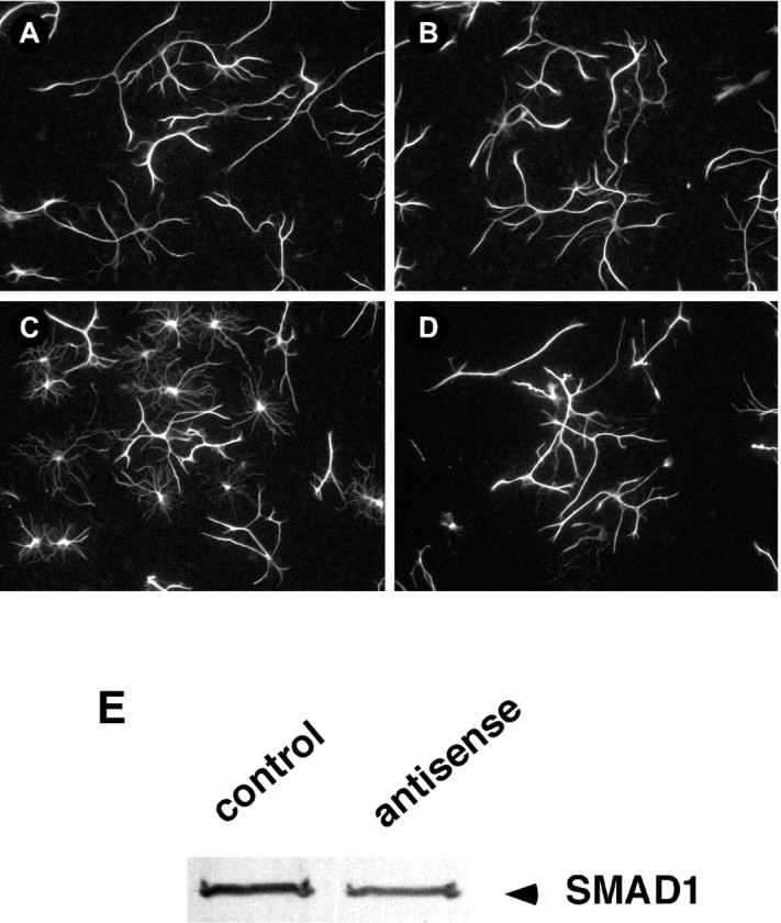
Previous studies have demonstrated that bone morphogenetic proteins (BMPs) activate the Smad1 signaling pathway to regulate cell determination and differentiation in the embryonic nervous system. Studies examining gene and protein expression in the rat cerebellum suggest that this pathway also regulates postnatal differentiation. Using microarrays, we found that Smad1 mRNA expression in the cerebellum increases transiently at postnatal day 6 (P6). Immunohistochemistry and Western blots showed that Smad1 and BMP4 proteins are present in the cerebellum, and that their expression also changes postnatally. The proteins are detectable at P4-P6, a stage at which most cerebellar cells reside in the external germinal layer (EGL), where they extensively differentiate. The levels become maximal at P8-P10, when neurons begin to migrate from the EGL into their mature positions in the internal granule layer. In cerebellar cultures prepared at P6 or P10, BMP4 activates Smad1 signaling to modulate cell differentiation. Brief BMP4 application caused Smad1 translocation from the neuronal cytoplasm into the nucleus, where it is known to regulate transcription in association with Smad4. Longer BMP4 treatment promoted the differentiation of both neuronal and non-neuronal cells. By 3 d, neuronal processes appeared more fasciculated, and the level of synaptotagmin, a protein found in synaptic vesicles, increased. In addition, many astroglial cells became more branched and stellate in morphology. The BMP-induced changes were reduced by treatment with antisense oligonucleotides to Smad1 or Smad4. These findings in vivo and in culture suggest that BMP4 and Smad1 signaling participate in regulating postnatal cerebellar differentiation.


Figures

Fig. 1.
Fig. 1.
Smad1 and BMP4 protein expression are temporally regulated in the cerebellum. Protein levels increase during early postnatal ontogeny and decline in the adult. A, Representative immunoblot showing Smad1 expression in cerebellar tissue prepared from rats at the indicated postnatal and adult ages.B, Immunoblot of BMP4 expression in cerebellar tissues prepared at the indicated ages. Similar developmental profiles of Smad1 and BMP4 expression were observed in four independent experiments. This blot was stripped and reprobed for actin as a control for sample preparation and loading.
Fig. 2.
Fig. 2.
Smad1 and BMP4 are expressed in the EGL. EGL cell-enriched samples from animals of the indicated ages were probed for Smad1 (A) or BMP4 (B). The BMP4 blot was stripped and reprobed for actin as a loading control (B). The proteins were detected at all postnatal ages examined. Similar results were obtained in four independent experiments.
Fig. 3.
Fig. 3.
BMP4 is expressed in several cell populations in the postnatal cerebellum. Sections prepared from cerebella isolated from rats at P10 were stained for BMP4 (A, B, D) or GFAP (C, E). BMP4 is detected in the EGL, in Purkinje neurons (P), and in cerebellar fiber tracts (WM). Little overlap is detected in the distribution of BMP4 and GFAP. A, Low-magnification view of tissue stained for BMP4. Insets are viewed at higher magnification in B–E. B, D, Higher-power views of BMP staining in the EGL and cerebellar fiber tracts.C, E, GFAP staining in the same regions shown inB, D.
Fig. 4.
Fig. 4.
BMP4 treatment induces Smad1 translocation into the nucleus. A–D, Fluorescence images of Smad1 staining in cerebellar cells maintained 4 d in culture and incubated in the absence (A, C) or presence (B, D) of BMP4 (10 ng/ml) for 45 min. A, B, Light microscopic images of fields of control (A) and BMP-treated (B) cells. C, D, Projections of confocal images of Smad1 staining in control (C) and BMP-treated (D) cells. Note that the number of Smad1-like immunoreactive patches in nuclei increases after BMP treatment.
Fig. 5.
Fig. 5.
Smad4 staining in control and BMP4-treated cells. Fluorescence images of Smad4 staining in cerebellar cells maintained for 4 d and then incubated for 45 min in the absence (A, C) or presence (B, D) of BMP4 (10 ng/ml).A, B, Light microscopic images of fields of control (A) and BMP-treated (B) cells. C, D, Confocal micrographs of individual control (C) and BMP-treated (D) cells show that nuclear staining is only slightly increased.
Fig. 6.
Fig. 6.
BMP4 induces granule neuron differentiation in cerebellar cultures. A, B, Images of MAP staining in cerebellar cells plated at P6 and grown for 3 d in the absence (A) or presence (B) of 10 ng/ml BMP4. BMP treatment promoted neuronal process outgrowth and branching in a dose-dependent manner. Similar changes were observed in cultures prepared at P10. C, Representative immunoblots of synaptotagmin 1a and MAP in cells maintained in culture in the absence or presence of 10 ng/ml BMP4 for 3 d. The BMP-induced changes in neuronal differentiation observed with immunofluorescence were accompanied by increases in the levels of these neuronal proteins.
Fig. 7.
Fig. 7.
BMP4 promotes the differentiation of astrocytes in cerebellar cultures. A–D, Images of GFAP staining in cells prepared at P6 and grown for 3 d in the absence or presence of increasing concentrations of BMP4. Similar effects were observed in cultures prepared at P10. A, Control. B, A total of 1 ng/ml BMP4. C, A total of 5 ng/ml BMP4.D, A total of 10 ng/ml BMP4.
Fig. 8.
Fig. 8.
BMP4-induced astrocyte differentiation is inhibited by treatment with a Smad1 antisense ODN. A–D, Fluorescence images of GFAP staining in cerebellar cells plated at P6 and grown for 3 d in the absence or presence of BMP4 (10 ng/ml) and/or the antisense ODN (3.5 μg/ml). A, Control.B, Smad1 ODN-treated cells. C, BMP4-treated cells. D, BMP4- and Smad1 ODN-treated cells. The Smad1 ODN inhibited the BMP4-induced changes in morphology but had no effect by itself. E, Immunoblot of Smad1 expression in cells grown in the absence (control) or presence (antisense) of the Smad1 antisense ODN. The ODN caused similar decreases in Smad1 expression in four independent experiments.

References

    1. Akazawa C, Ishibashi M, Shimizu C, Nakanishi S, Kageyama R. A mammalian helix-loop-helix factor structurally related to the product of Drosophila proneural gene atonal is a positive transcriptional regulator expressed in the developing nervous system. J Biol Chem. 1995;270:8730–8738. - PubMed
    1. Alder J, Lee KJ, Jessell TM, Hatten ME. Generation of cerebellar granule neurons in vivo by transplantation of BMP-treated neural progenitor cells. Nat Neurosci. 1999;2:535–540. - PubMed
    1. Altman J, Bayer SA. Development of the cerebellar system in relation to its evolution, structure, and functions. CRC; Boca Raton, FL: 1997.
    1. Aruga J, Minowa O, Yaginuma H, Kuno J, Nagai T, Noda T, Mikoshiba K. Mouse Zic1 is involved in cerebellar development. J Neurosci. 1998;18:284–293. - PMC - PubMed
    1. Beattie CE, Siegel RE. Developmental cues modulate GABA receptor subunit mRNA expression in cultured cerebellar granule neurons. J Neurosci. 1993;13:1784–1792. - PMC - PubMed

Publication types

MeSH terms

LinkOut - more resources