. 2002 Oct-Dec;3(4):254-9.
doi: 10.3348/kjr.2002.3.4.254.

Idiopathic synovial osteochondromatosis of the hip: radiographic and MR appearances in 15 patients

Affiliations

Idiopathic synovial osteochondromatosis of the hip: radiographic and MR appearances in 15 patients

Sung Hyun Kim et al. Korean J Radiol. 2002 Oct-Dec.

Abstract

Objective: To evaluate the radiographic and MR appearance of idiopathic synovial osteochondromatosis of the hip.

Materials and methods: Radiographs and MR images of 15 patients with idiopathic synovial osteochondromatosis of the hip were assessed. The former were analysed in terms of the presence of 1) juxta-articular calcified and/ or ossified bodies, 2) osteophytes, 3) bone erosion, 4) juxta-articular osteopenia, and 5) joint space narrowing, while for the latter, analysis focused on 1) the configuration of intra-articular bodies, 2) bone erosion, 3) synovial thickening, 4) conglomeration of intra-articular bodies, and 5) extra-articular extension.

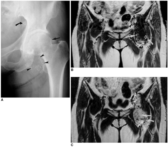
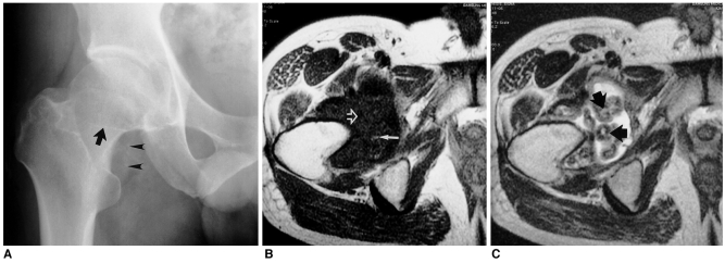
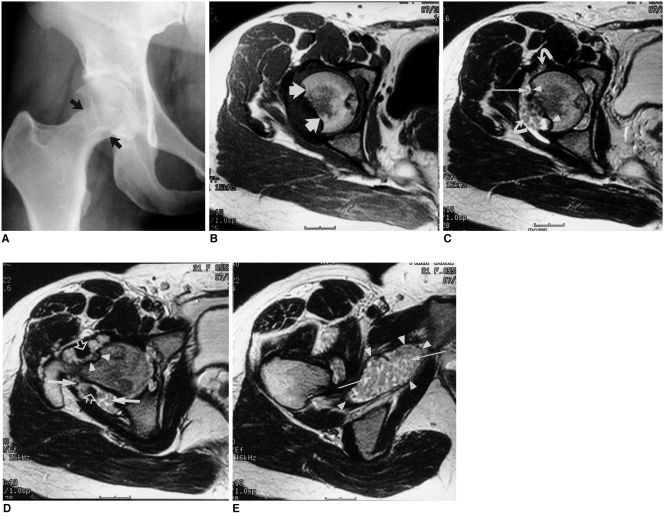
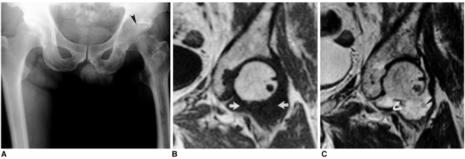
Results: At hip radiography, juxta-articular calcified and/ or ossified bodies were seen in 12 of the 15 patients (80%), bone erosion in eight (53%), osteophytes in seven (47%), juxta-articular osteopenia in five (33%) and joint space narrowing in five (33%). In eight patients (53%), MR imaging depicted intra-articular bodies of focal low signal intensity at all pulse sequences, and areas of isointensity at T1WI and hyperintensity at T2WI. In three (20%), intra-articular bodies of focal low signal intensity and areas of hyperintensity at all pulse sequences were observed, with areas of iso-intensity at T1WI and hyperintensity at T2WI, while in four (27%), intra-articular bodies of only focal low signal intensity at all pulse sequences were apparent. Synovial thickening was present in 13 patients (87%), bone erosion in 11 (73%), conglomeration of the intra-articular bodies in 11 (73%), and an extra-articular herniation sac in six (40%).

Conclusion: The most common radiographic finding of synovial osteochondromatosis of the hip was the presence of juxta-articular calcified and/ or ossified bodies. MR imaging depicted intra-articular bodies of focal low signal intensity at all pulse sequences, with areas of iso-intensity at T1WI and hyperintensity at T2WI. In addition, the presence of an extra-articular herniation sac was not uncommon.


Figures

Fig. 1
Fig. 1
A 52-year-old woman with idiopathic synovial osteochondromatosis of the left hip. A. Anteoposterior radiograph of the left hip depicts juxta-articular calicified and/ or ossified bodies (arrowheads), and an osteophyte (straight arrow). Note that superior to the iliopubic line, a calcified and/or ossified body (curved arrow) is also present, suggesting intrapelvic extension of the lesion. B. T1-weighted coronal MR image of the same patient shows an intra-articular lesion of low signal intensity (curved arrows), signal intensity which corresponds to an area of fat (straight arrow), and a conglomerated lesion (open arrow). Note that intrapelvic extension of the iliopsoas bursa has occurred (arrowheads). C. T2-weighted image, obtained at the same level as in B, shows intra-articular bodies of focal low signal intensity (curved arrows), and a conglomerated lesion (straight arrow). Note that intrapelvic extension of the iliopsoas bursa is again apparent (arrowheads).
Fig. 2
Fig. 2
A 37-year-old man with idiopathic synovial osteochondromatosis of the right hip. A. Anteoposterior radiograph of the right hip demonstrates the presence of juxta-articular calicified and/or ossified bodies (arrowheads), and an osteophyte (arrow). B. T1-weighted axial MR image of the same patient depicts intra-articular bodies of focal low signal intensity (open arrow) and areas of fat (straight arrow). C. T2-weighted image, obtained at the same level as in B, shows intra-articular bodies of focal low signal intensity and central areas of hyaline cartilage (arrows).
Fig. 3
Fig. 3
A 31-year-old woman with idiopathic synovial osteochondromatosis of the right hip. A. Anteoposterior radiograph of the right hip reveals extensive circumferential bone erosion and scalloping of the right femoral neck (arrows). B. T1-weighted axial MR image of the same patient depicts extensive bone erosion (arrows). C. T2-weighted image, obtained at the same level as in B, shows extensive bone erosion (arrowheads), intra-articular bodies without hyaline cartilage or fat (slender arrow), and an intra-articular body with central areas of hyaline cartilage (curved arrows). D. T2-weighted image of the same patient demonstrates extensive bone erosion (arrowheads) around the femoral neck, and the presence of intra-articular bodies (straight arrows). Note that thickening of the synovium (open arrows) has also occurred. E. T2-weighted image of the same patient shows an extra-articular herniation sac (arrowheads) filled with intra-articular bodies (arrows).
Fig. 4
Fig. 4
A 59-year-old man with idiopathic synovial osteochondromatosis of the left hip. A. Anteoposterior radiograph of the left hip shows juxta-articular osteopenia and joint-space narrowing of the left hip joint (arrowhead). B. T1-weighted coronal MR image of the same patient depicts an iso-intense intra-articular lesion (arrows). C. T2-weighted image, obtained at the same level as in B, shows an intra-articular body of low signal intensity (curved arrow). Note that another intra-articular body has peripheral low signal intensity (arrowhead), and the central area has the signal characteristics of hyaline cartilage.

References

    1. Milgram JW. Synovial osteochondromatosis: a histopathological study of thirty cases. J Bone Joint Surg Am. 1977;59:792–801. - PubMed
    1. Kramer J, Recht M, Deely DM, et al. MR appearance of idiopathic synovial osteochondromatosis. J Comput Assist Tomogr. 1993;17:772–776. - PubMed
    1. Llauger J, Palmer J, Roson N, Bague S, Camins A, Cremades R. Nonseptic monoarthritis: imaging features with clinical and histopathologic correlation. RadioGraphics. 2000;20:S263–S278. - PubMed
    1. Norman A, Steiner GC. Bone erosion in synovial chondromatosis. Radiology. 1986;161:749–752. - PubMed
    1. Crotty JM, Monu JUV, Pope TL. Synovial osteochondromatosis. Radiol Clin North Am. 1996;34:327–342. - PubMed