

CD4+CD25+ suppressor lymphocytes in the circulation of patients immunized against melanoma antigens
- PMID: 12514432
- PMCID: PMC2410039
- DOI: 10.1097/00002371-200301000-00009
CD4+CD25+ suppressor lymphocytes in the circulation of patients immunized against melanoma antigens
Abstract
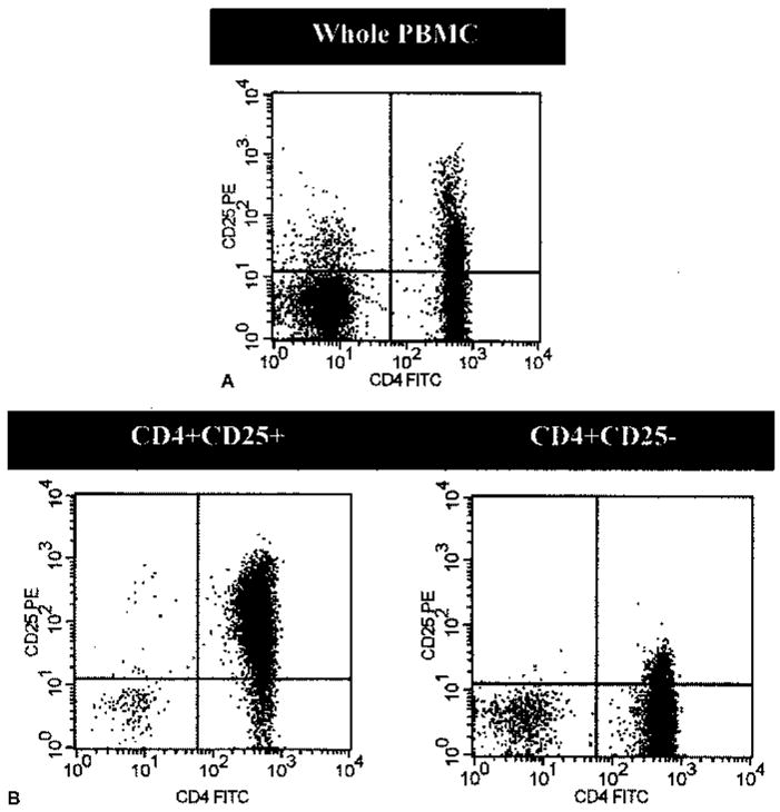
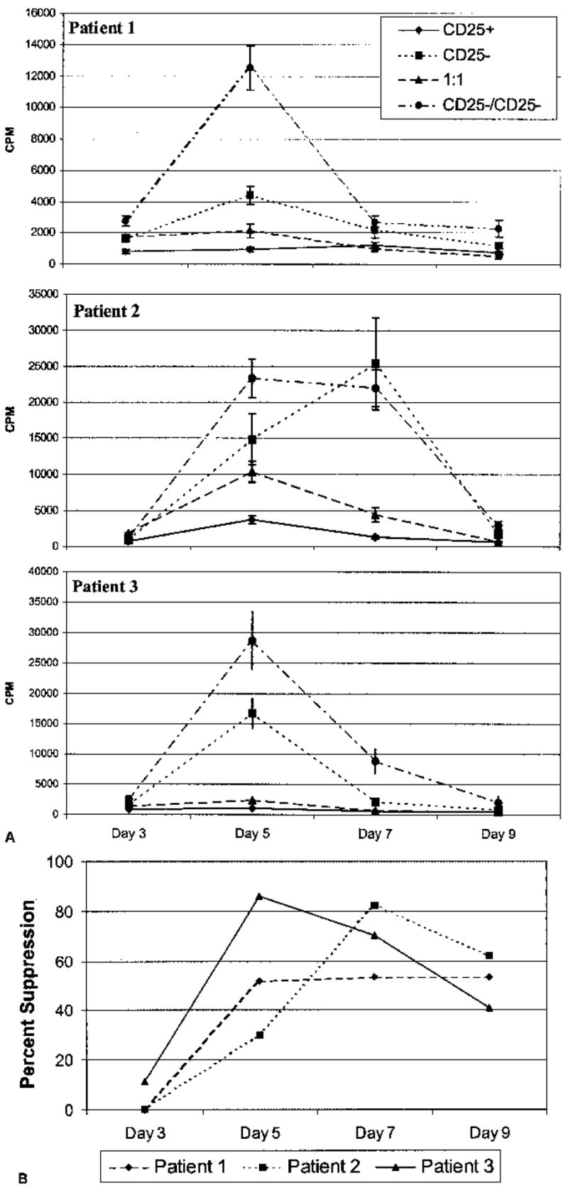
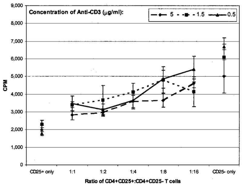
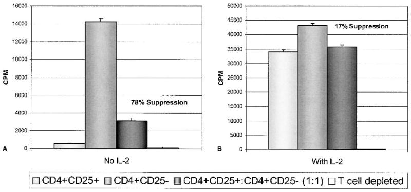
Murine studies have suggested that a population of CD4+ T cells expressing the alpha chain of the interleukin (IL)-2 receptor (CD25+) are phenotypically anergic in response to T cell receptor stimulation and can suppress the function of CD4+ and CD8+ T cells. Recent studies of peripheral lymphocytes from healthy human volunteers have identified a similar population, although little is known about the presence and activity of these cells in patients with cancer and their possible impact on anticancer immunization strategies. Thus, the authors have undertaken these studies in patients with metastatic melanoma undergoing immunizations with known melanoma antigens. CD4+ CD25+, CD4+ CD25-, and a 1:1 ratio of these isolated T cells were stimulated with soluble anti-CD3 antibody in the presence of irradiated T cell-depleted PBMCs, and proliferation was assessed by measuring [3H]thymidine incorporation. In 13 patients, isolated CD4+CD25+ T cells proliferated 68% (+/- 5.8%) less than separately cultured CD4+ CD25- T cells. Moreover, CD4+ CD25+ T cells suppressed the proliferation of an equal number of cocultured CD4+ CD25+ T cells in 11 of 13 patients by an average of 60% (+/- 4.9%). Suppression was not seen at day three of culture and became apparent at days five through nine. The degree of suppression was proportional to the numbers of CD4+ CD25+ T cells. Addition of high-dose IL-2 reversed the hypoproliferative phenotype of the CD4+ CD25+ T cells and abrogated their suppressive function. These studies demonstrate that anergic and functionally suppressive CD4+ CD25+ T cells exist in patients with melanoma undergoing tumor antigen immunization and thus may play a role in modifying the magnitude of the T cell response to immunization.
Figures
References
-
- Rosenberg SA. A new era for cancer immunotherapy based on the genes that encode cancer antigens. Immunity. 1999;10:281–7. - PubMed
-
- Rosenberg SA. Progress in human tumor immunology and immunotherapy. Nature. 2001;411:380–4. - PubMed
-
- Fu T, Shen Y, Fujimoto S. Tumor-specific CD4(+) suppressor T-cell clone capable of inhibiting rejection of syngeneic sarcoma in A/J mice. Int J Cancer. 2000;87:680–7. - PubMed
-
- Onizuka S, Tawara I, Shimizu J, et al. Tumor rejection by in vivo administration of anti-CD25 (interleukin-2 receptor alpha) monoclonal antibody. Cancer Res. 1999;59:3128–33. - PubMed
-
- Shimizu J, Yamazaki S, Sakaguchi S. Induction of tumor immunity by removing CD25+CD4+ T cells: a common basis between tumor immunity and autoimmunity. J Immunol. 1999;163:5211–8. - PubMed
MeSH terms
Substances
Grants and funding
LinkOut - more resources
Full Text Sources
Other Literature Sources
Medical
Research Materials
