. 2003 Jan 6;197(1):7-17.
doi: 10.1084/jem.20021229.

Mycobacteria target DC-SIGN to suppress dendritic cell function

Affiliations

Mycobacteria target DC-SIGN to suppress dendritic cell function

Teunis B H Geijtenbeek et al. J Exp Med. .

Abstract

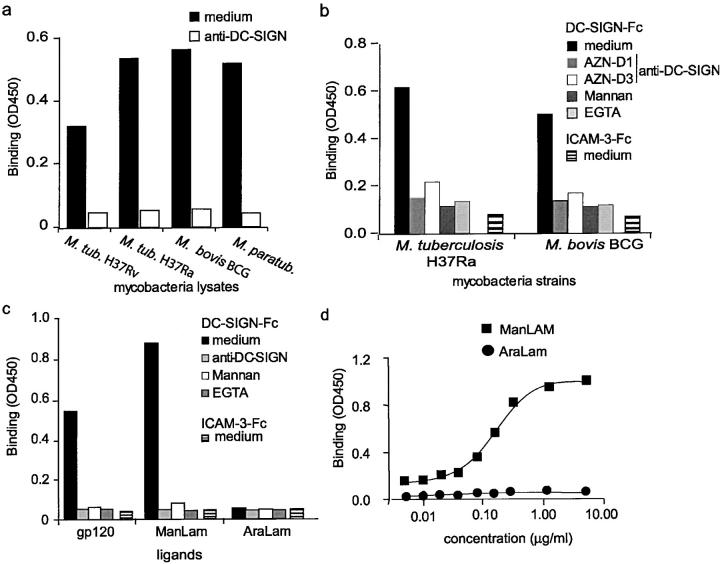
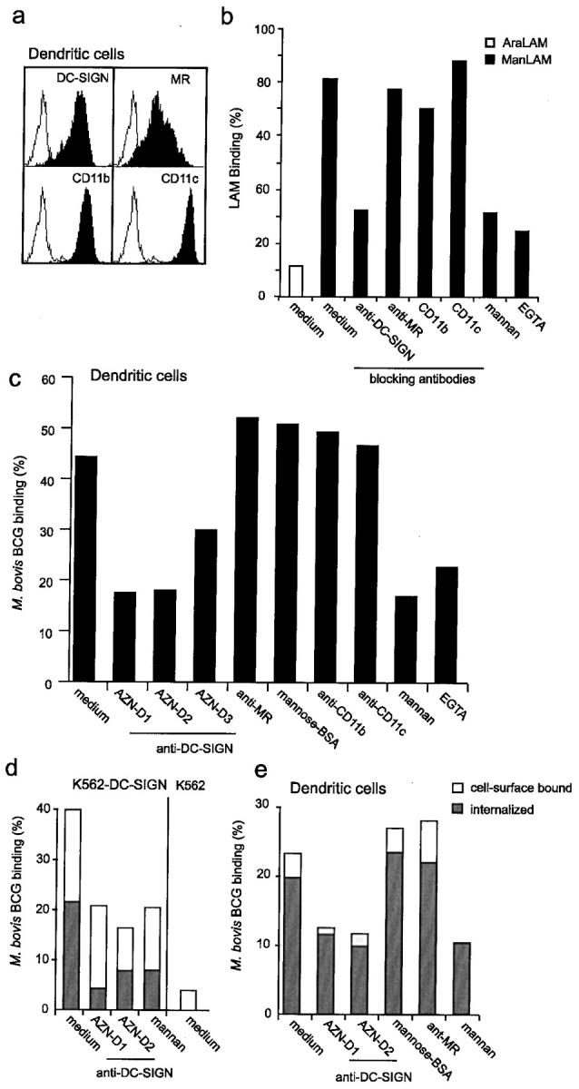
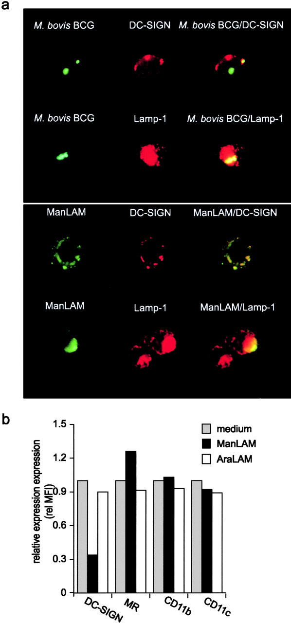
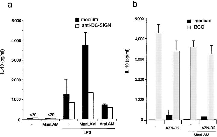
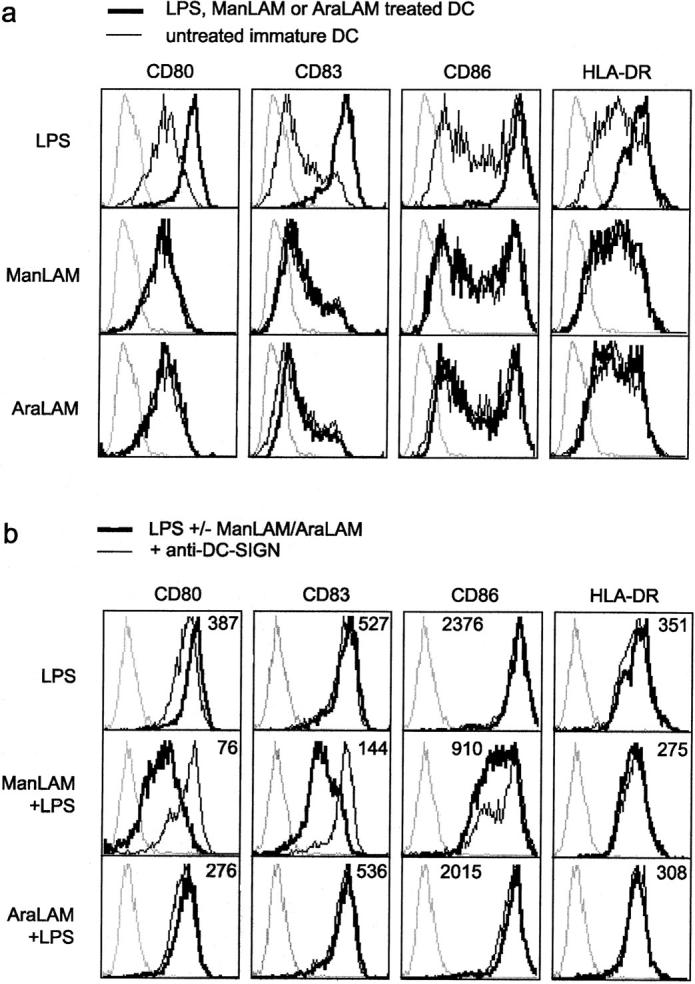
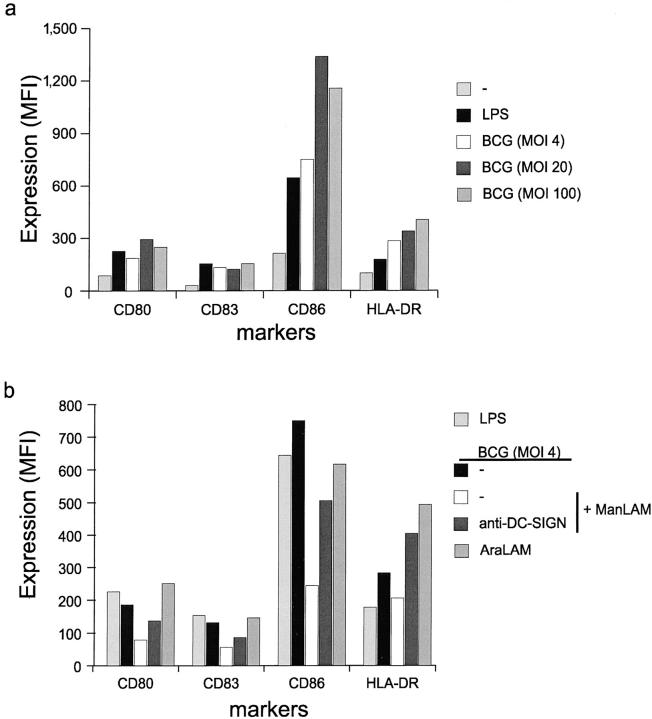
Mycobacterium tuberculosis represents a world-wide health risk and immunosuppression is a particular problem in M. tuberculosis infections. Although macrophages are primarily infected, dendritic cells (DCs) are important in inducing cellular immune responses against M. tuberculosis. We hypothesized that DCs represent a target for M. tuberculosis and that the observed immuno-suppression results from modulation of DC functions. We demonstrate that the DC-specific C-type lectin DC-SIGN is an important receptor on DCs that captures and internalizes intact Mycobacterium bovis bacillus Calmette-Guérin (BCG) through the mycobacterial cell wall component ManLAM. Antibodies against DC-SIGN block M. bovis BCG infection of DCs. ManLAM is also secreted by M. tuberculosis-infected macrophages and has been implicated as a virulence factor. Strikingly, ManLAM binding to DC-SIGN prevents mycobacteria- or LPS-induced DC maturation. Both mycobacteria and LPS induce DC maturation through Toll-like receptor (TLR) signaling, suggesting that DC-SIGN, upon binding of ManLAM, interferes with TLR-mediated signals. Blocking antibodies against DC-SIGN reverse the ManLAM-mediated immunosuppressive effects. Our results suggest that M. tuberculosis targets DC-SIGN both to infect DCs and to down-regulate DC-mediated immune responses. Moreover, we demonstrate that DC-SIGN has a broader pathogen recognition profile than previously shown, suggesting that DC-SIGN may represent a molecular target for clinical intervention in infections other than HIV-1.


Figures

Figure 1.
Figure 1.
DC-SIGN specifically binds ManLAM, a cell wall component of M. tuberculosis. (a) DC-SIGN interacts with several mycobacteria strains. DC-SIGN-Fc binding to mycobacteria (lysates; 5 μg) was determined by a Fc-specific ELISA. Specificity was determined by measuring binding in the presence of blocking DC-SIGN–specific antibodies (AZN-D1). Standard deviation <0.02 OD450. One representative experiment out of three is shown. (b) DC-SIGN interacts with viable mycobacteria strains. DC-SIGN-Fc binding to viable mycobacteria (5 × 10⁵ bacteria) was determined by a Fc-specific ELISA. Specificity was determined by measuring binding in the presence of blocking DC-SIGN–specific antibodies (AZN-D1 or AZN-D3) and mannan. EGTA was used to determine the calcium dependency of the DC-SIGN-Fc–mediated binding. ICAM-3-Fc binding to mycobacteria was also measured to exclude nonspecific binding by the Fc domain. Standard deviation <0.02 OD450. One representative experiment out of three is shown. (c) The mannosylated-lipoarabinomannan ManLAM, in contrast to the nonmannosylated AraLAM, is specifically bound by DC-SIGN. The anti–DC-SIGN antibody AZN-D1 was used to determine specificity. The DC-SIGN-Fc binding assay was performed as described for panel a. Standard deviation <0.02 OD450. One representative experiment out of three is shown. (d) DC-SIGN does not interact with AraLAM. The DC-SIGN-Fc binding assay was performed as described for panel a. One representative experiment out of three is shown.
Figure 2.
Figure 2.
Cellular DC-SIGN binds strongly to both viable mycobacteria and the mycobacterial component ManLAM through its primary binding site. (a) K562-DC-SIGN transfectants express high levels of DC-SIGN but lack expression of the other reported ManLAM receptors MR, CD11b, and CD11c. Transfectants were generated as described previously (reference 12). Open histograms represent the isotype controls, and filled histograms indicate the specific antibody staining. (b) DC-SIGN, expressed by K562 transfectants, binds strongly to intact M. bovis BCG and the mycobacterial component ManLAM but not to M. smegmatis and AraLAM. The adhesion of cells to the LAM glycans was determined using the fluorescent bead adhesion assay. Binding to viable mycobacteria was determined by measuring the binding of K562 transfectants to FITC-conjugated mycobacteria (MOI 20) using flow cytometry. Specificity was determined by measuring binding in the presence of blocking antibodies against DC-SIGN. Standard deviation for the fluorescent bead adhesion assay and the mycobacteria binding assay was <5 and <2%, respectively. One representative experiment out of three is shown. (c) The Val351 amino acid residue is not essential for the interaction of DC-SIGN with M. bovis BCG and ManLAM, similar to HIV-1 gp120, whereas it is essential for ICAM-3 binding. Binding to the V351G DC-SIGN mutant expressed by K562 cells was measured as described for panel b. Specificity was determined by measuring binding in the presence of blocking antibodies against DC-SIGN, mannan or EGTA. Standard deviation <5% (fluorescent bead adhesion assay) and <2% (mycobacteria binding assay). One representative experiment out of three is shown.
Figure 3.
Figure 3.
DC-SIGN is an important receptor for both ManLAM and mycobacteria on DCs. (a) Immature DCs express high levels of DC-SIGN and the other reported LAM receptors MR, CD11b, and CD11c. Open histograms represent isotype control and filled histograms indicate specific antibody staining. (b) Immature DCs bind strongly to ManLAM via DC-SIGN. Binding was determined using the fluorescent bead adhesion assay. Specificity was determined by measuring binding in the presence of mannan, EGTA or blocking antibodies against DC-SIGN (AZN-D2), MR (Clone 19), CD11b (bear-1), or CD11c (SHCL3). Standard deviation <5%. One representative experiment out of three is shown. (c) DC-SIGN mediates capture of M. bovis BCG by immature DCs. Binding was determined by flow cytometry using FITC-conjugated mycobacteria. Specificity was determined by measuring binding in the presence of antibodies against DC-SIGN (AZN-D1, AZN-D2, and AZN-D3), MR (Clone 19), CD11b (bear-1), and CD11c (SHCL3). Binding was also measured in the presence of the C-type lectin inhibitors mannan and EGTA, whereas a known MR ligand, mannose-BSA, was used to determine the contribution of the MR receptor. Standard deviation <2%. One representative experiment out of three is shown. (d) DC-SIGN mediates capture and internalization of M. bovis BCG by K562 cells. K562 transfectants were incubated with FITC-conjugated M. bovis BCG (MOI 20). Cells were washed, and surface FITC was quenched by exposure to trypan blue. Phagocytosis was determined by comparing the FITC emission before and after quenching using flow cytometry. Surface bound bacteria are represented by open bars, internalized by closed bars. Standard deviation <4%. One representative experiment out of three is shown. (e) Immature DCs rapidly phagocytose mycobacteria through DC-SIGN. The internalization was determined as described for panel b. Surface bound bacteria are represented by open bars, internalized by closed bars. Standard deviation <5%. One representative experiment out of three is shown.
Figure 4.
Figure 4.
DC-SIGN mediates internalization of captured mycobacteria and ManLAM. (a) M. bovis BCG and ManLAM are internalized by DC-SIGN on immature DCs and targeted to the lysosomes. The fate of captured mycobacteria was followed by analyzing immature DCs pulsed with FITC-conjugated M. bovis BCG (MOI 20) for 2 h using immunofluorescence microscopy (magnification 200×). ManLAM was followed by incubating DCs with ManLAM (10 μg/ml) for 1 h. DC-SIGN, ManLAM, and CD207a/LAMP-1 were stained with AZN-D1, F30.5, and H4A3, respectively. One representative experiment out of three is shown. (b) ManLAM induces down-regulation of DC-SIGN, but not of MR, CD11b, and CD11c. Immature DCs were incubated with 15 μg/ml of ManLAM or AraLAM for 18 h, and then DC-SIGN expression was determined by flow cytometry. One representative experiment out of three is shown.
Figure 5.
Figure 5.
Mycobacteria induce IL-10 production by DCs through ManLAM and direct infection. (a) ManLAM induces IL-10 production of LPS-matured DCs. Immature DCs were incubated with 15 μg/ml of either ManLAM or AraLAM in the presence of LPS (10 ng/ml). The specificity was determined in the presence of blocking antibodies against DC-SIGN (AZN-D2; 20 μg/ml). Supernatants were harvested after 18 h and the IL-10 production was measured by ELISA. Values are the means ± standard deviations of triplicate determinations. One representative experiment out of three is shown. (b) M. bovis BCG infection of immature DCs induces IL-10 production. Immature DCs were infected with M. bovis BCG (MOI 4), and the experiment was performed as described for panel a. Values are the means ± standard deviations of triplicate determinations. One representative experiment out of three is shown.
Figure 6.
Figure 6.
ManLAM inhibits LPS-induced DC activation through DC-SIGN binding. (a) ManLAM does not induce activation of immature DCs. Immature DCs were incubated with ManLAM, AraLAM, or LPS for 18 h, and activation was determined by measuring the expression of CD80, CD86, CD83, and HLA-DR. Dotted lines represent isotype controls, the thin lines indicate expression levels of immature DCs, and the thick line represents immature DCs that have been treated with either LPS (10 ng/ml), ManLAM (15 μg/ml), or AraLAM (15 μg/ml). One representative experiment out of three is shown. (b) LPS-induced activation of DCs is blocked by ManLAM. Immature DCs were cocultured with LPS alone, or together with either ManLAM or AraLAM for 18 h. Dotted lines represent isotype controls. Thick lines, and the mean fluorescence values in the histograms, represent the expression levels after treatment with LPS alone, or in combination with either ManLAM or AraLAM. Thin lines indicate the presence of antibodies against DC-SIGN throughout the incubation. One representative experiment out of three is shown.
Figure 7.
Figure 7.
M. bovis BCG induces maturation and ManLAM inhibits the induced DC activation through DC-SIGN binding. (a) M. bovis BCG induces maturation of immature DCs. Immature DCs were incubated with LPS or viable M. bovis BCG (MOI 4, 20, and 100) for 18 h, and activation was determined by measuring the expression of CD80, CD86, CD83, and HLA-DR. The mean fluorescence intensity (MFI) of the difference between specific antibody and isotype control is depicted. One representative experiment out of three is shown. (b) M. bovis BCG-induced activation of DCs is blocked by ManLAM. Immature DCs were infected with M. bovis BCG (MOI 4). Cells were preincubated with 15 μg/ml of either ManLAM or AraLAM and the expression of the markers was measured after 18 h as described for panel a. Specificity was determined by preincubating cells with blocking antibodies against DC-SIGN (AZN-D2; 20 μg/ml). One representative experiment out of three is shown.

Comment in

References

    1. Fenton, M.J., and M.W. Vermeulen. 1996. Immunopathology of tuberculosis: roles of macrophages and monocytes. Infect. Immun. 64:683–690. - PMC - PubMed
    1. Demangel, C., and W.J. Britton. 2000. Interaction of dendritic cells with mycobacteria: where the action starts. Immunol. Cell Biol. 78:318–324. - PubMed
    1. Banchereau, J., and R.M. Steinman. 1998. Dendritic cells and the control of immunity. Nature. 392:245–252. - PubMed
    1. Prigozy, T.I., P.A. Sieling, D. Clemens, P.L. Stewart, S.M. Behar, S.A. Porcelli, M.B. Brenner, R.L. Modlin, and M. Kronenberg. 1997. The mannose receptor delivers lipoglycan antigens to endosomes for presentation to T cells by CD1b molecules. Immunity. 6:187–197. - PubMed
    1. Manabe, Y.C., and W.R. Bishai. 2000. Latent Mycobacterium tuberculosis-persistence, patience, and winning by waiting. Nat. Med. 6:1327–1329. - PubMed

Publication types

MeSH terms