. 2003 Jan 7;100(1):38-43.
doi: 10.1073/pnas.0136947100. Epub 2002 Dec 23.

Crystal structure of saposin B reveals a dimeric shell for lipid binding

Affiliations

Crystal structure of saposin B reveals a dimeric shell for lipid binding

Victoria E Ahn et al. Proc Natl Acad Sci U S A. .

Abstract

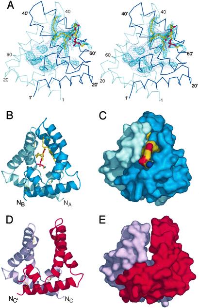
Saposin B is a small, nonenzymatic glycosphingolipid activator protein required for the breakdown of cerebroside sulfates (sulfatides) within the lysosome. The protein can extract target lipids from membranes, forming soluble protein-lipid complexes that are recognized by arylsulfatase A. The crystal structure of human saposin B reveals an unusual shell-like dimer consisting of a monolayer of alpha-helices enclosing a large hydrophobic cavity. Although the secondary structure of saposin B is similar to that of the known monomeric members of the saposin-like superfamily, the helices are repacked into a different tertiary arrangement to form the homodimer. A comparison of the two forms of the saposin B dimer suggests that extraction of target lipids from membranes involves a conformational change that facilitates access to the inner cavity.


Figures

Figure 1
Figure 1
(A) Multiple alignment of representative saposin-like proteins from eight different functional families (6). Sequence numbering is according to saposin B. The sequence alignment was generated with clustalw and includes manual adjustments. Identical amino acids are shaded black; conserved positions are shaded yellow. Experimentally determined N-glycosylation sites are indicated with red lettering. Human saposin B (SapB) is aligned with human saposins A, C, and D (SapA, SapC, and SapD), acyloxyacyl hydrolase (AOAH), plant phytepsin (PHY), acid sphingomyelinase (ASM), granulysin (NKG5), pore-forming amoebapore A (AP-A), Countinin (Count), surfactant-associated protein B (SP-B), and porcine NK-lysin (NKL). Leading and trailing dots indicate cases where the saposin motif is a sub-sequence of a larger protein. Note that in the phytepsin “swaposin” domain, the sequence block VVSQ… TFDG precedes the block ADPM… NRLP, as marked by the red asterisk (32). The secondary structures of saposin B chain A (Top) and NK-lysin (Bottom) are shown as cylinders representing helices, and the cysteines that are linked by disulfide bonds are connected with brackets. A turn of 310 helix at the end of helix α2 in saposin B is indicated by hatching. The saposin B residues are assigned to the following environment classes: residues contributing >20 Å2 to the dimer interface (circles); residues facing the inner cavity but not part of the dimer interface (squares); residues with side chains lining the inner hydrophobic cavity (black symbols); residues with side chain lining the opening to the cavity (white symbols). The inner cavity is formed only in the AB dimer. (BD) Ribbon diagrams of chains A and B of saposin B and of the NK-lysin monomer, respectively. The side chains from chain A that are exposed to the inner cavity are indicated in stick representation.
Figure 2
Figure 2
The saposin dimer encloses a large hydrophobic cavity. (A) Stereo view of the AB dimer. The Cα trace of the A chain is in light blue, and the B chain is in dark blue. Residues from the B chain are labeled with primes. The surface of the inner cavity is in gray mesh, and the positive Fo-Fc density calculated before the inclusion of the ligand is indicated in dark blue mesh (contoured at 1.5 σ). The final refined phosphatidylethanolamine fragment is in stick representation. (B and C) Ribbon and solvent-accessible surface diagrams of the AB dimer, with coloring as in A. The lipid headgroup is exposed to solvent, and the acyl chains are contained within the cavity. Disulfide bonds are indicated with thin yellow lines. (D and E) Ribbon and surface representations of the symmetric CC′ dimer with the chains in pink and red. There are tight interchain contacts at the base, but no contacts between the hairpin regions and no ordered ligands within the solvent-exposed core.
Figure 3
Figure 3
(A) Surface burial in the dimer for each of the residues in the three crystallographically independent chains. The open “front” (B) and sealed “back” (C) of the AB dimer are shown. The lipid headgroup protrudes from the opening to the cavity in the front, whereas contacts among His-31(B), Y50(A), and Y54(A) seal the cavity on the back. (D) The relative orientations of residues H31(C), Y50(C′), and Y54(C′) (and the equivalent residues related by the twofold symmetry, not shown in this view) in the CC′ dimer are similar to those found near the opening to the AB dimer.
Figure 4
Figure 4
(A) Superposition of the AB and CC′ dimers, shown in tube representation. Chains A (light blue) and C (pink) superimpose well, as do the base of chains B (dark blue) and C′ (red). The kink in the third helix of chain B is evident in the overlay with the C′ chain. Residues Met-10 (base) and Ile-46 (hairpin) are near the molecular (pseudo)-symmetry axes and are indicated in black. Equivalent residues in the dimers are connected by black lines. (B) SDS/PAGE of reduced and nonreduced saposin B with and without the additional cysteines. Wild-type saposin B is in lanes 1 and 4, the M10C mutant is in lanes 2 and 5, and I46C is in lanes 3 and 6. DTT was added to lanes 1–3 only. (C) Activation of cerebroside sulfate hydrolysis by arylsulfatase A in the presence of wild-type recombinant saposin B (squares), M10C (circles), and I46C (triangles) crosslinked dimers. Less than 2% hydrolysis of the input sulfatide was observed in the absence of activator under the assay conditions.

References

    1. Sandhoff K, Kolter T. Trends Cell Biol. 1996;6:98–103. - PubMed
    1. Vaccaro A M, Salvioli R, Tatti M, Ciaffoni F. Neurochem Res. 1999;24:307–314. - PubMed
    1. Fluharty A L. Trends Glycosci Glycotechnol. 1995;7:167–189.
    1. Wilkening G, Linke T, Uhlhorn-Dierks G, Sandhoff K. J Biol Chem. 2000;275:35814–35819. - PubMed
    1. Munford R S, Sheppard P O, O'Hara P J. J Lipid Res. 1995;36:1653–1663. - PubMed

Publication types

Associated data

LinkOut - more resources