

Imaging whole Escherichia coli bacteria by using single-particle x-ray diffraction
- PMID: 12518059
- PMCID: PMC140897
- DOI: 10.1073/pnas.232691299
Imaging whole Escherichia coli bacteria by using single-particle x-ray diffraction
Abstract
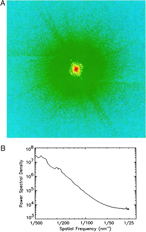
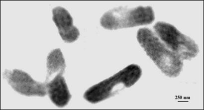
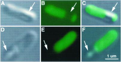
We report the first experimental recording, to our knowledge, of the diffraction pattern from intact Escherichia coli bacteria using coherent x-rays with a wavelength of 2 A. By using the oversampling phasing method, a real space image at a resolution of 30 nm was directly reconstructed from the diffraction pattern. An R factor used for characterizing the quality of the reconstruction was in the range of 5%, which demonstrated the reliability of the reconstruction process. The distribution of proteins inside the bacteria labeled with manganese oxide has been identified and this distribution confirmed by fluorescence microscopy images. Compared with lens-based microscopy, this diffraction-based imaging approach can examine thicker samples, such as whole cultured cells, in three dimensions with resolution limited only by radiation damage. Looking forward, the successful recording and reconstruction of diffraction patterns from biological samples reported here represent an important step toward the potential of imaging single biomolecules at near-atomic resolution by combining single-particle diffraction with x-ray free electron lasers.
Figures
References
-
- Sayre D. In: Imaging Processes and Coherence in Physics. Springer Lecture Notes in Physics. Schlenker M, editor. Vol. 112. Berlin: Springer; 1980. pp. 229–235.
-
- Miao J, Charalambous P, Kirz J, Sayre D. Nature. 1999;400:342–344.
-
- Robinson I K, Vartanyants I A, Williams G J, Pferfer M A, Pitney J A. Phys Rev Lett. 2001;87:195505. - PubMed
-
- Miao J, Ishikawa T, Johnson B, Anderson E H, Lai B, Hodgson K O. Phys Rev Lett. 2002;89:088303. - PubMed
-
- Miao J, Ohsuna T, Terasaki O, Hodgson K O, O'Keefe M A. Phys Rev Lett. 2002;89:155502. - PubMed
Publication types
MeSH terms
LinkOut - more resources
Full Text Sources
Other Literature Sources
