. 2003 Jan 7;100(1):137-42.
doi: 10.1073/pnas.0135800100. Epub 2002 Dec 23.

Asymmetry of the central apparatus defines the location of active microtubule sliding in Chlamydomonas flagella

Affiliations

Asymmetry of the central apparatus defines the location of active microtubule sliding in Chlamydomonas flagella

Matthew J Wargo et al. Proc Natl Acad Sci U S A. .

Abstract

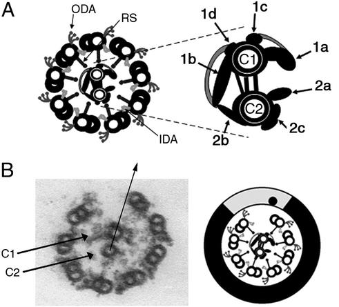
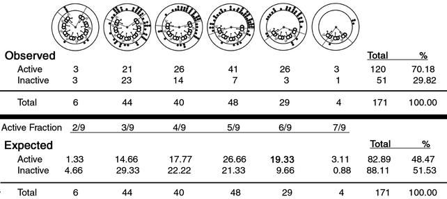
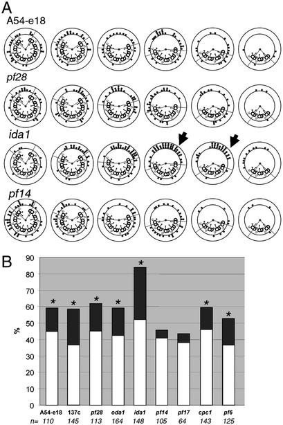
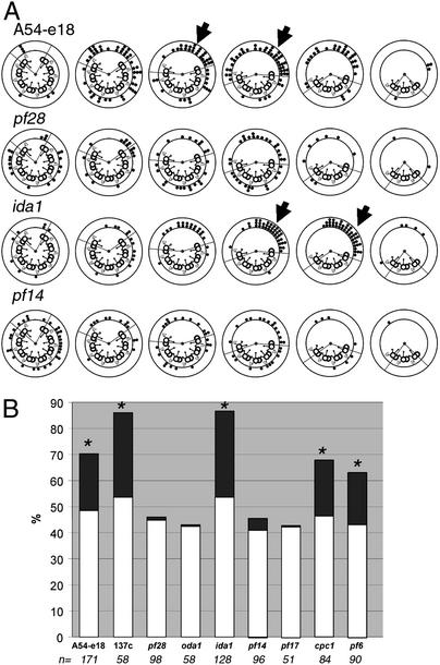
Regulation of ciliary and flagellar motility requires spatial control of dynein-driven microtubule sliding. However, the mechanism for regulating the location and symmetry of dynein activity is not understood. One hypothesis is that the asymmetrically organized central apparatus, through interactions with the radial spokes, transmits a signal to regulate dynein-driven microtubule sliding between subsets of doublet microtubules. Based on this model, we hypothesized that the orientation of the central apparatus defines positions of active microtubule sliding required to control bending in the axoneme. To test this, we induced microtubule sliding in axonemes isolated from wild-type and mutant Chlamydomonas cells, and then used electron microscopy to determine the orientation of the central apparatus. Transverse sections of wild-type axonemes revealed that the C1 microtubule is predominantly oriented toward the position of active microtubule sliding. In contrast, the central apparatus is randomly oriented in axonemes isolated from radial spoke deficient mutants. For outer arm dynein mutants, the C1 microtubule is oriented toward the position of active microtubule sliding in low calcium buffer, but is randomly oriented in high calcium buffer. These results provide evidence that the central apparatus defines the position of active microtubule sliding, and may regulate the size and shape of axonemal bends through interactions with the radial spokes. In addition, our results indicate that in high calcium conditions required to generate symmetric waveforms, the outer dynein arms are potential targets of the central pair-radial spoke control system.


Figures

Figure 1
Figure 1
(A) Transverse section of flagellar axoneme. The outer and inner dynein arms (ODA, IDA) and radial spokes (RS) are attached to the outer doublet microtubules. The central apparatus is enlarged and offset. Central pair projections are labeled according to Mitchell and Sale (10). (B) Electron micrograph and accompanying diagram, axoneme transverse section after microtubule sliding. The C1 and C2 microtubules of the central pair are labeled. The arrow defines the orientation of the central pair. The point at which the arrow intersects the circumference of the doublets is indicated by a dot on the accompanying diagram. The position of active microtubule sliding includes the area lightly shaded. The inactive area includes the remainder of the axoneme (dark shading).
Figure 2
Figure 2
Quantitative analysis of central apparatus orientation. The example shown is wild-type axonemes. We calculated the expected number of events for both the active and inactive areas for each pattern of microtubule sliding, if the orientation of the central apparatus was random. Expected events = (fraction of the axoneme in the active/inactive area) × (total events for sliding pattern). The expected events in the active area were totaled for all sliding patterns (Total), expressed as a percentage of the total number of transverse sections (%), and compared with the percentage of observed events.
Figure 3
Figure 3
Central apparatus orientation, low-calcium buffer. (A) Examples of data obtained from axonemes isolated from four strains after microtubule sliding. The lines drawn from the center of each cross section delineate the active and inactive areas defined in Fig. 1B. The dots represent the orientation of the C1 microtubule for individual transverse sections. Particularly striking examples of the C1 microtubule oriented toward the position of microtubule sliding are indicated by arrows. To view all data, see Fig. 6. (B) Percent of transverse sections in which the C1 microtubule was oriented toward the active area. Light bars indicate the expected percentage of events. The difference between the expected and observed percentages is darkly shaded. Strains in which this difference is significant are indicated by asterisks (χ2 test; P < 0.03). The total number of transverse sections included is indicated below each mutant.
Figure 4
Figure 4
Central apparatus orientation, high calcium buffer. (A) Examples of data obtained from axonemes isolated from four strains after microtubule sliding in pCa4 buffer. The lines drawn from the center of each cross section delineate the active and inactive areas defined in Fig. 1B. The dots represent the orientation of the C1 microtubule for individual transverse sections. Particularly striking examples of the C1 microtubule oriented toward the position of microtubule sliding are indicated by arrows. To view all data, see Fig. 6. (B) Percentage of transverse sections in which the C1 microtubule was oriented toward the active area as shown in A. Light bars indicate the expected percentage of events. The difference between the expected and observed values is darkly shaded. Strains in which this difference is significant are indicated by asterisks (χ2 test; P < 0.01). The total number of transverse sections included is indicated below each mutant.
Figure 5
Figure 5
As the central apparatus rotates clockwise, the C1 microtubule contacts specific radial spokes, which in turn relay a regulatory signal to the dynein arms on specific subsets of doublet microtubules. The doublets and associated dynein arms actively engaged in microtubule sliding are indicated by light shading. In each subsequent transverse section (left to right), microtubule sliding is visualized as progressive loss of doublet microtubules from the axoneme.

References

    1. Satir P. J Cell Biol. 1968;26:805–834. - PMC - PubMed
    1. Summers K, Gibbons I R. Proc Natl Acad Sci USA. 1971;68:3092–3096. - PMC - PubMed
    1. Brokaw C J. Science. 1972;178:455–462. - PubMed
    1. Shingyoji C, Murakami A, Takahashi K. Nature. 1977;265:269–270. - PubMed
    1. Brokaw C J. Science. 1989;243:1593–1596. - PubMed

Publication types