. 2003 Feb;18(2):90-9.
doi: 10.1002/hbm.10078.

Dorsal penile nerve stimulation elicits left-hemisphere dominant activation in the second somatosensory cortex

Affiliations

Dorsal penile nerve stimulation elicits left-hemisphere dominant activation in the second somatosensory cortex

J P Mäkelä et al. Hum Brain Mapp. 2003 Feb.

Abstract

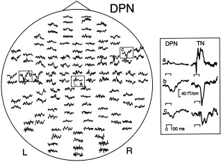
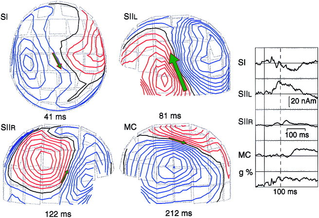
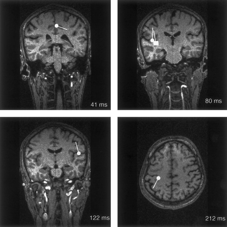
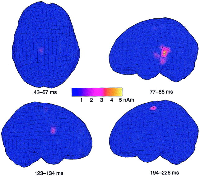
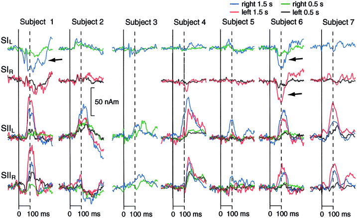
Activation of peripheral mixed and cutaneous nerves activates a distributed cortical network including the second somatosensory cortex (SII) in the parietal operculum. SII activation has not been previously reported in the stimulation of the dorsal penile nerve (DPN). We recorded somatosensory evoked fields (SEFs) to DPN stimulation from 7 healthy adults with a 122-channel whole-scalp neuromagnetometer. Electrical pulses were applied once every 0.5 or 1.5 sec to the left and right DPN. For comparison, left and right median and tibial nerves were stimulated alternatingly at 1.5-sec intervals. DPN stimuli elicited weak, early responses in the vicinity of responses to tibial nerve stimulation in the primary somatosensory cortex. Strong later responses, peaking at 107-126 msec were evoked in the SII cortices of both hemispheres, with left-hemisphere dominance. In addition to tactile processing, SII could also contribute to mediating emotional effects of DPN stimuli.


Figures

Figure 1
Figure 1
Responses to left‐sided DPN stimuli in Subject 2 (ISI 1.5 sec, 200 responses averaged). Each trace shows a signal detected by one sensor as a function of time, and signals obtained by orthogonal planar gradiometers from the same location are shown as pairs. The responses have been digitally filtered with a 0.03–90 Hz passband. The head is viewed from above and the nose points upwards. The scale bars in the encircled area (a) indicate 100 msec and 20 fT/cm. Inset (right): Enlarged responses from the SI (a) and left (b) and right (c) SII regions. Responses to left‐sided TN stimuli from the same regions are depicted for comparison.
Figure 2
Figure 2
Multidipole model for responses to left‐sided DPN stimuli (ISI 1.5 sec) of Subject 2. In addition to SI and bilateral SII signals, a prolonged activation is evident in the motor cortex (MC). Insert (right): Dipole strength as a function of time; the vertical dashed line indicates time point 100 msec after the stimulus. The step in isocontour plots is 10 fT. The dipole moment and goodness‐of fit (g) values are 10 nAm and 83% at 41 msec, 25 nAm and 92% at 81 msec, 10 nAm and 82% at 122 msec, and 9 nAm and 93% at 212 msec.
Figure 3
Figure 3
Locations of sources of responses to left DPN stimuli at 0.5‐sec ISI in the longitudinal fissure (41 msec) and to left DPN stimuli at 1.5‐sec ISI in the SII cortices (80 and 122 msec) and in the motor cortex (212 msec) in Subject 2, superimposed on his MR image. The square in the left SII (top right) indicates source location at 80 msec, and the circle the source at 112 msec.
Figure 4
Figure 4
Minimum current estimates of responses to left‐sided DPN stimuli (ISI 1.5 sec) in Subject 2. The areas modeled with current dipoles in Figure 2 show MCE activity at corresponding latencies.
Figure 5
Figure 5
Responses to DPN stimuli presented at ISIs of 0.5 and 1.5 sec in all subjects. The strongest signals of each individual from SI and SII regions are displayed. The arrows point to deflections generated in the nearby posterior parietal cortex.

References

    1. Adolphs R, Damasio H, Tranel D, Cooper G, Damasio A (2000): A role for somatosensory cortices in the visual recognition of emotion as revealed by three‐dimensional lesion mapping. J Neurosci 20: 2683–2690. - PMC - PubMed
    1. Ahonen A, Hämäläinen M, Kajola M, Knuutila J, Laine P, Lounasmaa O, Parkkonen L, Simola J, Tesche C (1993): 122‐channel SQUID instrument for investigating the magnetic signals from human brain. Phys Scripta T49: 198–205.
    1. Alary F, Simoes C, Jousmäki V, Forss N, Hari R (2002): Cortical activity associated with passive movements of human index finger: a MEG study. NeuroImage 15: 691–696. - PubMed
    1. Allison T, McCarthy G, Wood C, Williamson P, Spencer D (1989): Human cortical potentials evoked by stimulation of the median nerve. II. Cytoarchitechtonic areas generating long‐latency activation. J Neurophysiol 62: 711–722. - PubMed
    1. Allison T, McCarthy G, Luby M, Puce A, Spencer D (1996): Localization of functional regions of human mesial cortex by somatosensory evoked potential recording and by cortical stimulation. Electroenceph Clin Neurophysiol 100: 126–140. - PubMed

LinkOut - more resources