

Alterations in T-cell receptor Vbeta repertoire of CD4 and CD8 T lymphocytes in human immunodeficiency virus-infected children
- PMID: 12522039
- PMCID: PMC145262
- DOI: 10.1128/cdli.10.1.53-58.2003
Alterations in T-cell receptor Vbeta repertoire of CD4 and CD8 T lymphocytes in human immunodeficiency virus-infected children
Abstract
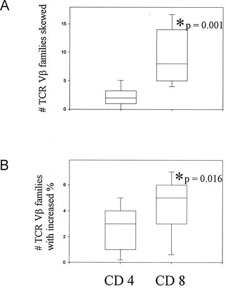
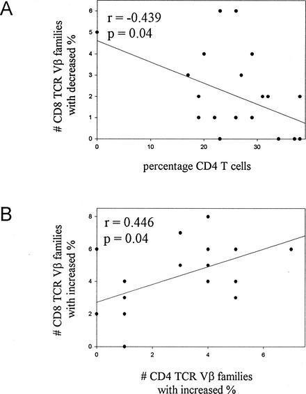
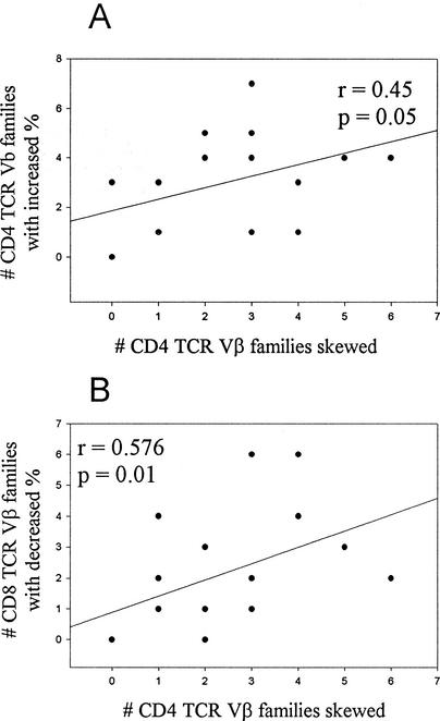
Perturbations in the T-cell receptor (TCR) Vbeta repertoire were assessed in the CD4 and CD8 T lymphocytes of human immunodeficiency virus (HIV)-infected children who were receiving therapy during the chronic phase of infection by flow cytometry (FC) and PCR analysis. By FC, representation of 21 TCR Vbeta subfamilies was assessed for an increased or decreased percentage in CD4 and CD8 T cells, and by PCR, 22 TCR Vbeta subfamilies of CD4 and CD8 T cells were analyzed by CDR3 spectratyping for perturbations and reduction in the number of peaks, loss of Gaussian distribution, or clonal dominance. The majority of the TCR Vbeta subfamilies were examined by both methods and assessed for deviation from the norm by comparison with cord blood samples. The CD8-T-lymphocyte population exhibited more perturbations than the CD4 subset, and clonal dominance was present exclusively in CD8 T cells. Of the 55 total CD8-TCR Vbeta families classified with clonal dominance by CDR3 spectratyping, only 18 of these exhibited increased expression by FC. Patients with high numbers of CD8-TCR Vbeta families with decreased percentages had reduced percentages of total CD4 T cells. Increases in the number of CD4-TCR Vbeta families with increased percentages showed a positive correlation with skewing. Overall, changes from normal were often discordant between the two methods. This study suggests that the assessment of HIV-induced alterations in TCR Vbeta families at cellular and molecular levels yields different information and that our understanding of the immune response to HIV is still evolving.
Figures
Similar articles
-
CD4+ and CD8+ T cell receptor repertoire perturbations with normal levels of T cell receptor excision circles in HIV-infected, therapy-naive adolescents.AIDS Res Hum Retroviruses. 2003 Jun;19(6):487-95. doi: 10.1089/088922203766774531. AIDS Res Hum Retroviruses. 2003. PMID: 12882658
-
Antiretroviral therapy restores diversity in the T-cell receptor Vbeta repertoire of CD4 T-cell subpopulations among human immunodeficiency virus type 1-infected children and adolescents.Clin Vaccine Immunol. 2009 Sep;16(9):1293-301. doi: 10.1128/CVI.00074-09. Epub 2009 Jul 15. Clin Vaccine Immunol. 2009. PMID: 19605599 Free PMC article.
-
T-Cell receptor Vbeta repertoire CDR3 length diversity differs within CD45RA and CD45RO T-cell subsets in healthy and human immunodeficiency virus-infected children.Clin Diagn Lab Immunol. 2000 Nov;7(6):953-9. doi: 10.1128/CDLI.7.6.953-959.2000. Clin Diagn Lab Immunol. 2000. PMID: 11063505 Free PMC article.
-
T-cell receptor repertoire in healthy Sardinian subjects.Hum Immunol. 2003 Jul;64(7):689-95. doi: 10.1016/s0198-8859(03)00086-7. Hum Immunol. 2003. PMID: 12826371
-
Tracking phenotypically and functionally distinct T cell subsets via T cell repertoire diversity.Mol Immunol. 2008 Feb;45(3):607-18. doi: 10.1016/j.molimm.2006.05.017. Epub 2007 Aug 24. Mol Immunol. 2008. PMID: 17719639 Free PMC article. Review.
Cited by
-
Perinatal clinical course of Vici syndrome associated with novel EPG5 variants: unique cardiac changes and difficulty with foetal diagnosis.BMJ Case Rep. 2024 Jan 5;17(1):e255847. doi: 10.1136/bcr-2023-255847. BMJ Case Rep. 2024. PMID: 38182173
-
Distribution and evolution of T-cell receptor Vbeta repertoire on peripheral blood lymphocytes of newborn infants of human immunodeficiency virus (HIV)-infected mothers: differential display on CD4 and CD8 T cells and effect of HIV infection.Clin Vaccine Immunol. 2007 Sep;14(9):1215-22. doi: 10.1128/CVI.00092-07. Epub 2007 Jul 25. Clin Vaccine Immunol. 2007. PMID: 17652526 Free PMC article.
-
T-cell receptor phenotype pattern in atopic children using commercial fluorescently labeled antibodies against 21 human class-specific v segments for the tcrβ chain (vβ) of peripheral blood: a cross sectional study.Allergy Asthma Clin Immunol. 2016 Mar 2;12:10. doi: 10.1186/s13223-016-0115-3. eCollection 2016. Allergy Asthma Clin Immunol. 2016. PMID: 26941803 Free PMC article.
-
Significantly skewed memory CD8+ T cell subsets in HIV-1 infected infants during the first year of life.Clin Immunol. 2009 Mar;130(3):280-9. doi: 10.1016/j.clim.2008.09.006. Epub 2008 Nov 8. Clin Immunol. 2009. PMID: 18996749 Free PMC article.
-
Dynamic Perturbations of the T-Cell Receptor Repertoire in Chronic HIV Infection and following Antiretroviral Therapy.Front Immunol. 2016 Jan 11;6:644. doi: 10.3389/fimmu.2015.00644. eCollection 2015. Front Immunol. 2016. PMID: 26793190 Free PMC article.
References
-
- Blattman, J. N., D. J. D. Sourdive, K. Murali-Krishna, R. Ahmed, and J. D. Altman. 2000. Evolution of the T cell repertoire during primary, memory, and recall responses to viral infection. J. Immunol. 165:6081-6090. - PubMed
-
- Callan, M. F. C., N. Steven, P. Krausa, J. D. K. Wilson, P. A. H. Moss, G. M. Gillespie, J. I. Bell, A. B. Rickinson, and A. J. McMichael. 1996. Large clonal expansions of CD8 T cells in acute infectious mononucleosis. Nat. Med. 2:906-911. - PubMed
-
- Connors, M., J. A. Kovacs, S. Krevat, J. C. Gea-Banacloche, M. C. Sneller, M. Flanigan, J. A. Metcalf, R. E. Walker, J. Falloon, M. Baseler, R. Stevens, I. Feuerstein, H. Masur, and H. C. Lane. 1997. HIV induces changes in CD4+ T-cell phenotype and depletions within the CD4+ T-cell repertoire that are not immediately restored by antiviral or immune-based therapies. Nat. Med. 3:533-540. - PubMed
-
- Cossarizza, A., C. Ortolani, C. Mussini, G. Guaraldi, N. Mongiardo, V. Borghi, D. Barbieri, E. Bellesia, M. G. Franseschini, B. D. Rienzo, and C. Franceschi. 1995. Lack of selective Vβ deletion in CD4 or CD8 T lymphocytes and functional integrity of TCR during acute HIV syndrome. AIDS 9:547-553. - PubMed
-
- aDouek, D. C., J. M. Brenchley, M. R. Betts, D. R. Ambrozak, B. J. Hill, Y. Okamoto, J. P. Casazza, J. Kuruppu, K. Kunstman, S. Wolinsky, Z. Grossman, M. Dybul, A. Oxenius, D. A. Price, M. Connors, and R. A. Koup. 2002. HIV preferentially infects HIV-specific CD4+ T cells. Nature 417:95-98. - PubMed
Publication types
MeSH terms
Substances
Grants and funding
LinkOut - more resources
Full Text Sources
Medical
Research Materials
