. 2003 Jan;10(1):140-53.
doi: 10.1128/cdli.10.1.140-153.2003.

Functional and morphological development of lymphoid tissues and immune regulatory and effector function in rhesus monkeys: cytokine-secreting cells, immunoglobulin-secreting cells, and CD5(+) B-1 cells appear early in fetal development

Affiliations

Functional and morphological development of lymphoid tissues and immune regulatory and effector function in rhesus monkeys: cytokine-secreting cells, immunoglobulin-secreting cells, and CD5(+) B-1 cells appear early in fetal development

Norbert Makori et al. Clin Diagn Lab Immunol. 2003 Jan.

Abstract

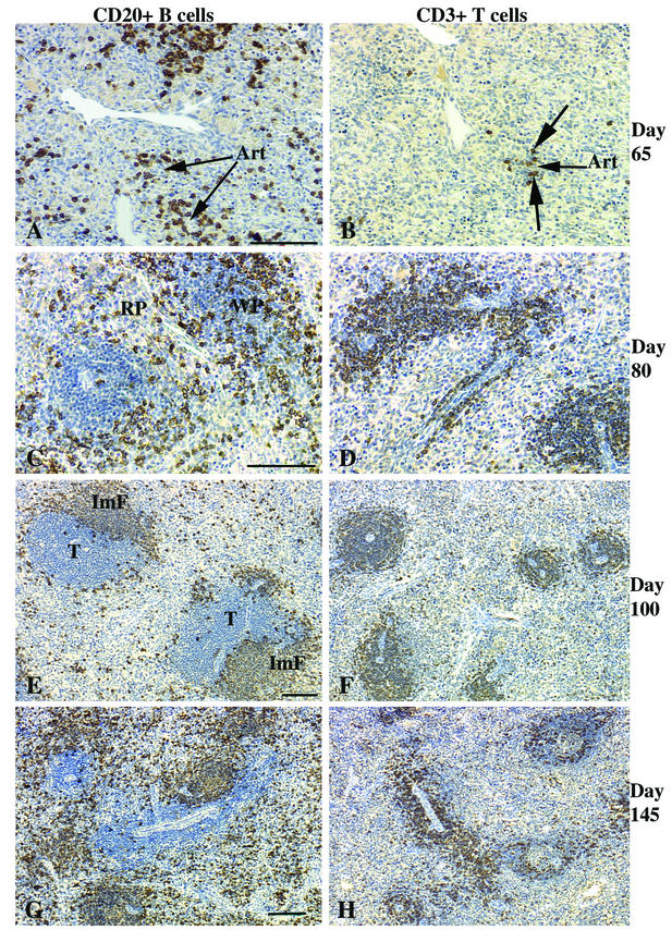
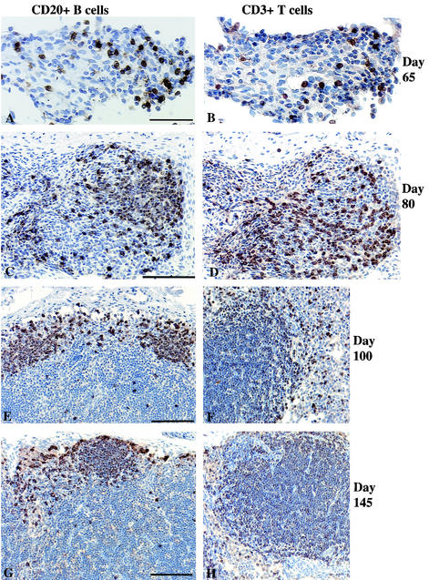
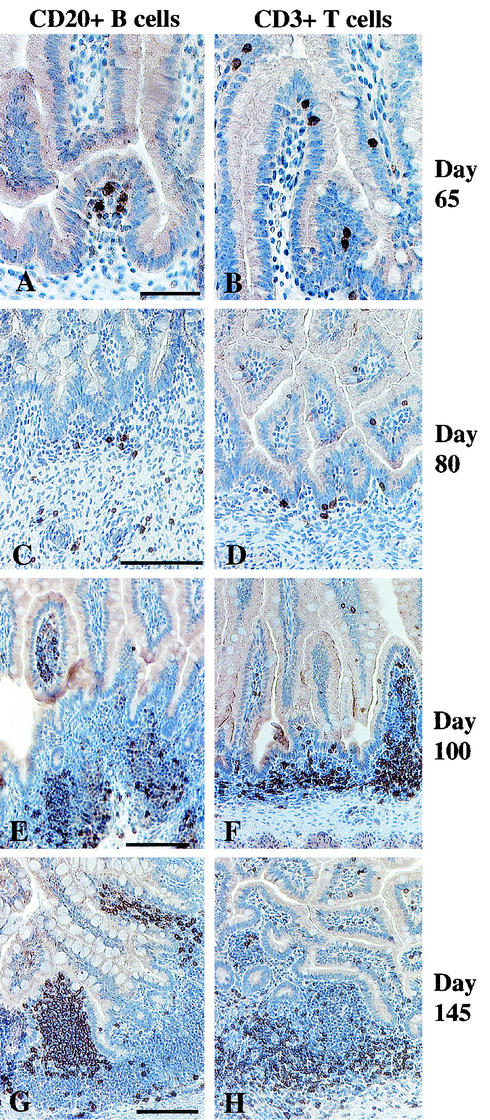
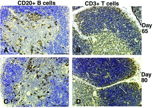
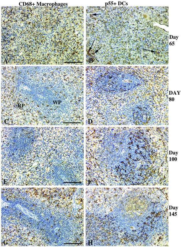
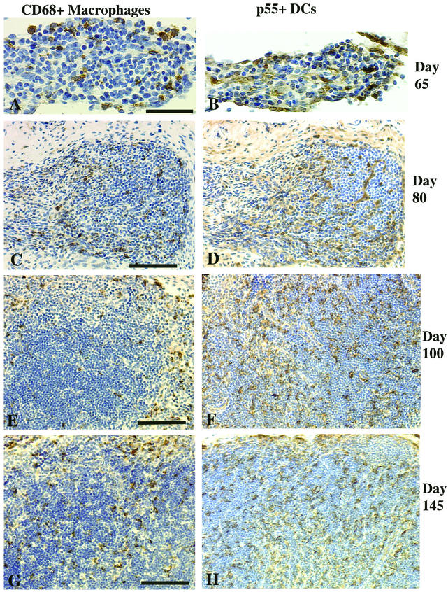
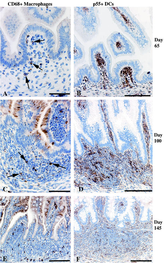
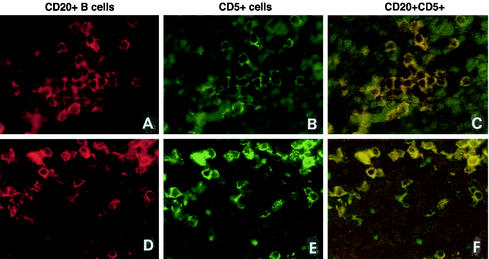
Little is known regarding the timing of immune ontogeny and effector function in fetal humans and nonhuman primates. We studied the organization of lymphocyte and antigen-presenting cell populations in developing lymphoid tissues of rhesus monkey fetuses during the second and third trimesters (65 to 145 days of gestation; term = 165 days). Immunoglobulin-secreting and cytokine-secreting cells were detected at day 80. The thymus, spleen, lymph nodes, and intestinal mucosa were examined for cells expressing CD3, CD5, CD20, CD68, p55, and HLA-DR. In the spleens of 65-day-old fetuses (early second trimester), the overwhelming majority of total lymphocytes were CD5(+) CD20(+) B-1 cells. The remaining lymphocytes were CD3(+) T cells. By day 80, splenic B and T cells were equal in number. Intraepithelial CD3(+) CD5(-) T cells and lamina propria CD20(+) CD5(+) B cells were present in the intestines of 65-day-old fetuses. By day 80, numerous CD20(+) CD5(+) B cells were present in the jejunums and colons and early lymphocyte aggregate formation was evident. The spleens of 80- to 145-day-old fetuses contained immunoglobulin M (IgM)-secreting cells, while IgA-, IgG-, interleukin-6-, and gamma interferon-secreting cells were numerous in the spleens and colons. Thus, by the second trimester, the lymphoid tissues of the rhesus monkey fetus have a complete repertoire of properly organized antigen-presenting cells, T cells, and B cells.


Figures

FIG.1.
FIG.1.
Spleens of fetal rhesus monkeys. At 65 days of gestation, >90% of the lymphocytes of the rhesus monkey fetus are B cells (A) with a few T cells (B). Note that some B-cell clusters and all T cells are associated with the ends of arterioles (Art). No distinguishable WP areas were apparent at day 65. (C and D) By day 80, immature WP areas (1:1 ratio of B to T cells) were distinct, with many B cells at the edge of the forming T-cell compartment. (E and F) Immature follicles (ImF in panel E) in the outer margins of the WP T-cell compartment (T) at day 100 of gestation. Note that at days 80 (D) and 100 (F), >80% of the T cells are in the WP, with the remainder scattered in the RP. The WP increased in size compared to the RP by day 145. (G) B cells; (H) T cells. Bars, 100 μm.
FIG.2.
FIG.2.
Mesenteric lymph nodes of fetal rhesus monkeys. At 65 days of gestation, a small number of B cells (A) and T cells (B) in a ratio of 1:1 were scattered among large numbers of mesenchymal cells. At 80 days of gestation, large numbers of B cells (C) and T cells (D) showed evidence of early organization into distinct T- and B-cell compartments. At day 100 of gestation, B cells (E) formed a thin layer outside the central T-cell area (F). At day 145 of gestation, B-cell follicles (G) were well defined, as was the T-cell compartment in the paracortex (H). The axillary lymph nodes had developmental features similar to those of the mesenteric lymph nodes, but axillary lymph node development was relatively delayed at day 65. Bars, 50 μm (A) and 100 μm (C, E, and G).
FIG. 3.
FIG. 3.
Jejunums of fetal rhesus monkeys. At 65 days of gestation, B cells in the lamina propria (A) and scattered T cells mainly in the intraepithelial compartment (B); at 80 days of gestation, B-cell clusters (C) and individual T cells (D) in the lamina propria; (E to H) well-defined lymphoid aggregates in the lamina propria at day 100 (E and F) and day 145 (G and H) of gestation; (E and G) B cells on the luminal side of the aggregates; (F and H) widespread T cells in the lamina propria and concentrations of T cells on the muscularis side of the lymphoid aggregates. Bars, 50 μm (A) and 100 μm (C, E, and G).
FIG. 4.
FIG. 4.
Thymuses of fetal rhesus monkeys. (A and B) At day 65 of gestation, B cells can be seen throughout the corticomedullary junction (A) and intensely stained CD3+ thymocytes can be seen in the cortex and medulla (B). (C and D) Similar cell populations were observed at day 80 and in older fetuses (data not shown). Bars, 100 μm.
FIG. 5.
FIG. 5.
Spleens of fetal rhesus monkeys. At day 65 of gestation, CD68+ macrophages (A) and p55+ DCs (arrows) (B); at day 80 of gestation (C and D) and day 100 of gestation (E and F), macrophages in the RP with a few scattered in the WP (C and E); (D and F) DCs located within the WP T-cell areas; (G) at day 145 of gestation, macrophages common in the WP; (H) DCs mainly in T-cell areas. Bars, 100 μm.
FIG. 6.
FIG. 6.
Mesenteric lymph nodes of fetal rhesus monkeys. At day 65 of gestation, scattered macrophages (A) and a few DCs (B) can be seen. At day 80 of gestation, the macrophage (C) and DC (D) populations had increased. At day 100 (E) and day 145 (G) of gestation, macrophages were concentrated in the outer cortex, while at day 100 (F) and day 145 (H) of gestation, the DCs were mainly confined to the paracortex. Bars, 50 μm (A) and 100 μm (C, E, and G).
FIG. 7.
FIG. 7.
Jejunums, ileums, and colons of fetal rhesus monkeys. (A and B) At day 65 of gestation, macrophages were few (A; arrows), while p55+ DCs were numerous (B) throughout the gut lamina propria, especially at the tips of villi. (C and D) In lymphoid aggregates at day 100 of gestation, macrophages were rare in the T-cell areas (C; arrows), while DCs were numerous (D). (E) At day 145 of gestation, macrophages were numerous within the lymphoid aggregates. (F) DCs were located in T-cell areas. Bars, 50 μm (A and C) and 100 μm (B to F).
FIG. 8.
FIG. 8.
Fetal CD20+ B cells express CD5. Double immunofluorescence with anti-CD20 (A and D; Texas red) and anti-CD5 (B and E; FITC green) in the spleen (A, B, and C) and mesenteric lymph nodes (D, E, and F) at day 65 of gestation shows double-stained cells. The majority of the CD20+ B cells in all lymphoid tissues at days 65 to 145 of gestation expressed CD5. Double-labeled cells are yellow, CD5+ single-labeled positive cells are green, and CD20+ single-labeled positive cells are red. Magnification, ×340.

Similar articles

Cited by

References

    1. Ahluwalia, B., B. Wesley, O. Adeyiga, D. M. Smith, A. Da-Silva, and S. Rajguru. 2000. Alcohol modulates cytokine secretion and synthesis in human fetus: an in vivo and in vitro study. Alcohol 21:207-213. - PubMed
    1. Attanasio, R., J. S. Allan, S. A. Anderson, T. C. Chanh, and R. C. Kennedy. 1991. Anti-idiotypic antibody response to monoclonal anti-CD4 preparations in nonhuman primate species. J. Immunol. 146:507-514. - PubMed
    1. Bals, R., C. Lang, D. J. Weiner, C. Vogelmeier, U. Welsch, and J. M. Wilson. 2001. Rhesus monkey (Macaca mulatta) mucosal antimicrobial peptides are close homologues of human molecules. Clin. Diagn. Lab. Immunol. 8:370-375. - PMC - PubMed
    1. Baumgarth, N., O. C. Herman, G. C. Jager, L. Brown, and L. A. Herzenberg. 1999. Innate and acquired humoral immunities to influenza virus are mediated by distinct arms of the immune system. Proc. Natl. Acad. Sci. USA 96:2250-2255. - PMC - PubMed
    1. Bhat, N. M., A. B. Kantor, M. M. Bieber, A. M. Stall, L. A. Herzenberg, and N. N. Teng. 1992. The ontogeny and functional characteristics of human B-1 (CD5+ B) cells. Int. Immunol. 4:243-252. - PubMed

Publication types

MeSH terms