

Involvement of tachykinin receptors in Clostridium perfringens beta-toxin-induced plasma extravasation
- PMID: 12522069
- PMCID: PMC1573648
- DOI: 10.1038/sj.bjp.0705022
Involvement of tachykinin receptors in Clostridium perfringens beta-toxin-induced plasma extravasation
Abstract
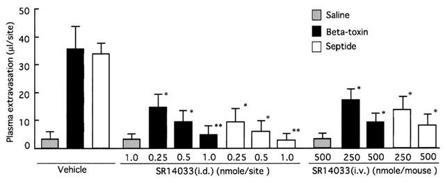
1 Clostridium perfringens beta-toxin causes dermonecrosis and oedema in the dorsal skin of animals. In the present study, we investigated the mechanisms of oedema induced by the toxin. 2 The toxin induced plasma extravasation in the dorsal skin of Balb/c mice. 3 The extravasation was significantly inhibited by diphenhydramine, a histamine 1 receptor antagonist. However, the toxin did not cause the release of histamine from mouse mastocytoma cells. 4 Tachykinin NK(1) receptor antagonists, [D-Pro(2), D-Trp(7,9)]-SP, [D-Pro(4), D-Trp(7,9)]-SP and spantide, inhibited the toxin-induced leakage in a dose-dependent manner. Furthermore, the non-peptide tachykinin NK(1) receptor antagonist, SR140333, markedly inhibited the toxin-induced leakage. 5 The leakage induced by the toxin was markedly reduced in capsaicin-pretreated mouse skin but the leakage was not affected by systemic pretreatment with a calcitonin gene-related peptide receptor antagonist (CGRP(8-37)). 6 The toxin-induced leakage was significantly inhibited by the N-type Ca(2+) channel blocker, omega-conotoxin MVIIA, and the bradykinin B(2) receptor antagonist, HOE140 (D-Arg-[Hyp(3), Thi(5), D-Tic(7), Oic(8)]-bradykinin), but was not affected by the selective L-type Ca(2+) channel blocker, verapamil, the P-type Ca(2+) channel blocker, omega-agatoxin IVA, tetrodotoxin (TTX), the TTX-resistant Na(+) channel blocker, carbamazepine, or the sensory nerve conduction blocker, lignocaine. 7 These results suggest that plasma extravasation induced by beta-toxin in mouse skin is mediated via a mechanism involving tachykinin NK(1) receptors.
Figures
Similar articles
-
Involvement of vanilloid receptors and purinoceptors in the Phoneutria nigriventer spider venom-induced plasma extravasation in rat skin.Eur J Pharmacol. 2000 Mar 17;391(3):305-15. doi: 10.1016/s0014-2999(00)00075-3. Eur J Pharmacol. 2000. PMID: 10729373
-
Involvement of tumour necrosis factor-alpha in Clostridium perfringens beta-toxin-induced plasma extravasation in mice.Br J Pharmacol. 2008 Mar;153(6):1296-302. doi: 10.1038/bjp.2008.9. Epub 2008 Feb 11. Br J Pharmacol. 2008. PMID: 18264118 Free PMC article.
-
Characterization of the receptor and the mechanisms underlying the inflammatory response induced by des-Arg9-BK in mouse pleurisy.Br J Pharmacol. 1998 Jan;123(2):281-91. doi: 10.1038/sj.bjp.0701590. Br J Pharmacol. 1998. PMID: 9489617 Free PMC article.
-
Characterization of the mechanisms underlying the inflammatory response to Polistes lanio lanio (paper wasp) venom in mouse dorsal skin.Toxicon. 2009 Jan;53(1):42-52. doi: 10.1016/j.toxicon.2008.10.006. Epub 2008 Oct 17. Toxicon. 2009. PMID: 18977380
-
Tachykinin receptors antagonists: from research to clinic.Curr Drug Targets. 2006 Aug;7(8):975-92. doi: 10.2174/138945006778019381. Curr Drug Targets. 2006. PMID: 16918326 Review.
Cited by
-
Clostridium Perfringens Toxins Involved in Mammalian Veterinary Diseases.Open Toxinology J. 2010;2:24-42. Open Toxinology J. 2010. PMID: 24511335 Free PMC article.
-
Recent progress in understanding the pathogenesis of Clostridium perfringens type C infections.Vet Microbiol. 2011 Nov 21;153(1-2):37-43. doi: 10.1016/j.vetmic.2011.02.048. Epub 2011 Feb 26. Vet Microbiol. 2011. PMID: 21420802 Free PMC article. Review.
-
Molecular Evolutionary Constraints that Determine the Avirulence State of Clostridium botulinum C2 Toxin.J Mol Evol. 2017 Apr;84(4):174-186. doi: 10.1007/s00239-017-9791-y. Epub 2017 Apr 5. J Mol Evol. 2017. PMID: 28382496
-
Blood proteome profiling for biomarker discovery in broilers with necrotic enteritis.Sci Rep. 2025 Apr 15;15(1):12895. doi: 10.1038/s41598-025-97783-w. Sci Rep. 2025. PMID: 40234672 Free PMC article.
-
Mechanisms of Action and Cell Death Associated with Clostridium perfringens Toxins.Toxins (Basel). 2018 May 22;10(5):212. doi: 10.3390/toxins10050212. Toxins (Basel). 2018. PMID: 29786671 Free PMC article. Review.
References
-
- AKOPIAN A.N., SIVILOTTI L., WOOD J.N. A tetrodotoxin-resistant voltage-gated sodium channel expressed by sensory neurons. Nature. 1996;379:257–262. - PubMed
-
- ALBER G., SCHEUBER P.H., RECK B., SAILER-KRAMER B., HARTMANN A., HAMMER D.K. Role of substance P in immediate-type skin reactions induced by staphylococcal enterotoxin B in unsensitized monkeys. J. Allergy. Clin. Immunol. 1989;84:880–885. - PubMed
-
- ARBUCKLE J.B., DOCHERTY R.J. Expression of tetrodotoxin-resistant sodium channels in capsaicin-sensitive dorsal root ganglion neurons of adult rats. Neurosci. Lett. 1995;185:70–73. - PubMed
-
- BACCEI M.L., KOCSIS J.D. Voltage-gated calcium currents in axotomized adult rat cutaneous afferent neurons. J. Neurophysiol. 2000;83:2227–2238. - PubMed
Publication types
MeSH terms
Substances
LinkOut - more resources
Full Text Sources
Other Literature Sources
Medical
Research Materials
Miscellaneous
