. 2003 Jan 21;100(2):663-8.
doi: 10.1073/pnas.242728499. Epub 2003 Jan 8.

WNK1, a kinase mutated in inherited hypertension with hyperkalemia, localizes to diverse Cl- -transporting epithelia

Affiliations

WNK1, a kinase mutated in inherited hypertension with hyperkalemia, localizes to diverse Cl- -transporting epithelia

Keith A Choate et al. Proc Natl Acad Sci U S A. .

Abstract

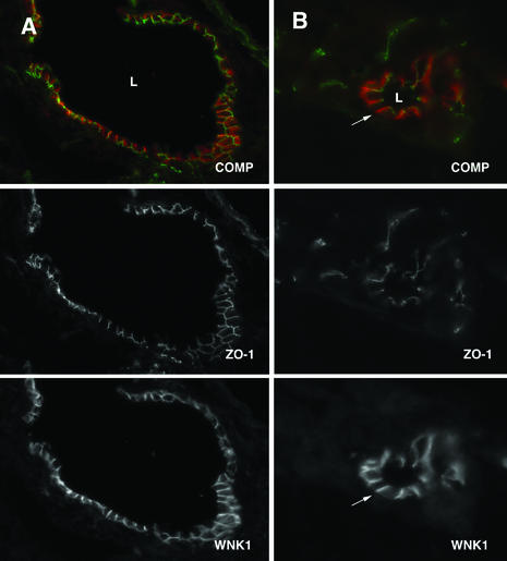
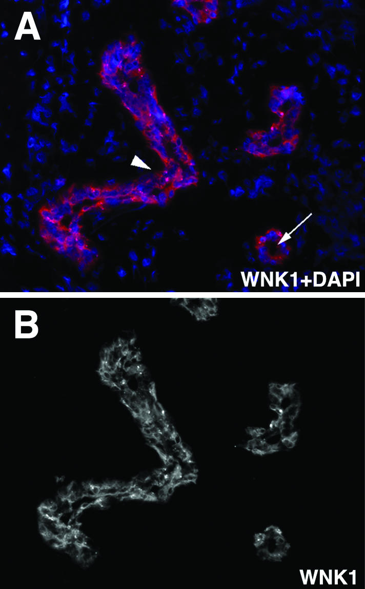
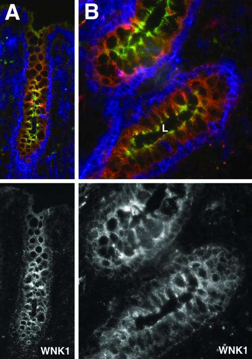
Mutations in WNK1 and WNK4, genes encoding members of a novel family of serine-threonine kinases, have recently been shown to cause pseudohypoaldosteronism type II (PHAII), an autosomal dominant disorder featuring hypertension, hyperkalemia, and renal tubular acidosis. The localization of these kinases in the distal nephron and the Cl(-) dependence of these phenotypes suggest that these mutations increase renal Cl(-) reabsorption. Although WNK4 expression is limited to the kidney, WNK1 is expressed in many tissues. We have examined the distribution of WNK1 in these extrarenal tissues. Immunostaining using WNK1-specific antibodies demonstrated that WNK1 is not present in all cell types; rather, it is predominantly localized in polarized epithelia, including those lining the lumen of the hepatic biliary ducts, pancreatic ducts, epididymis, sweat ducts, colonic crypts, and gallbladder. WNK1 is also found in the basal layers of epidermis and throughout the esophageal epithelium. The subcellular localization of WNK1 varies among these epithelia. WNK1 is cytoplasmic in kidney, colon, gallbladder, sweat duct, skin, and esophagus; in contrast, it localizes to the lateral membrane in bile ducts, pancreatic ducts, and epididymis. These epithelia are all notable for their prominent role in Cl(-) flux. Moreover, these sites largely coincide with those involved in the pathology of cystic fibrosis, a disease characterized by deranged epithelial Cl(-) flux. Together with the known pathophysiology of PHAII, these findings suggest that WNK1 plays a general role in the regulation of epithelial Cl(-) flux, a finding that suggests the potential of new approaches to the selective modulation of these processes.


Figures

Figure 1
Figure 1
Northern analysis of mouse WNK1 transcripts. A mouse WNK1 cDNA probe orthologous to exons 12–14 of human WNK1 (GenBank accession no. NM_018979) was hybridized to RNA from a mouse multiple tissue Northern blot (CLONTECH). WNK1 transcripts are most strongly expressed in kidney, heart, and testis. Two transcripts of ≈8 and 10 kb are observed. The 8-kb isoform is predominant in kidney, whereas the 10-kb isoform is the major transcript in other tissues. The positions of the RNA size standards in kilobases are indicated.
Figure 2
Figure 2
WNK1 immunolocalization in hepatic bile duct epithelium. Frozen sections of mouse liver were stained with anti-WNK1 (red) and anti-ZO-1 (green) antibody as described in Methods and analyzed by fluorescence microscopy. WNK1 is localized to cholangiocytes, cuboidal epithelial cells that line the biliary tract lumen. (A) A large intrahepatic bile duct. (Top) Composite (COMP) staining for WNK1 and ZO-1. (Middle and Bottom) Staining with each antibody alone. (B) Demonstration of WNK1 localization in a smaller bile ductule. As in A, WNK1 is localized to the lateral membrane (demonstrated by the arrow), whereas ZO-1 is localized to the tight junction. L, bile duct lumen.
Figure 3
Figure 3
Expression of WNK1 in exocrine pancreatic duct epithelium. Frozen sections of mouse pancreas were stained with anti-WNK1 (red) and anti-ZO-1 antibody (green) and analyzed by fluorescence microscopy as in Fig. 2. A longitudinal section of pancreatic ducts is shown. WNK1 is present in the lateral membrane of cuboidal epithelial cells of large main exocrine pancreatic ducts (arrows). WNK1 is not expressed in smaller intralobular ducts (arrowheads).
Figure 4
Figure 4
WNK1 immunolocalization in epididymal epithelium. Frozen sections of mouse epididymis were stained with anti-WNK1 (red) and anti-ZO-1 antibody (green) and analyzed by fluorescence microscopy (AC) and confocal microscopy (DF). (A) WNK1 expression is seen in the columnar epithelial cells of the epididymis. WNK1 becomes more tightly membrane associated with lateral accentuation with progression from basal cell layers (Ba) to superficial layers (Su) of this pseudostratified epithelium. (B) Same view as in A, with ZO-1 staining alone. (C) Same view as in A, with WNK1 staining alone. (D) Confocal composite of WNK1 and ZO-1 demonstrating the lateral localization of WNK1 in epididymal epithelium. (E) Same view as in D, showing ZO-1 staining alone. (F) Same view as in D, showing WNK1 staining alone. Arrow demonstrates the lateral expression of WNK1, beginning at the tight junction.
Figure 5
Figure 5
Localization of WNK1 in gallbladder epithelium. Frozen sections of mouse gallbladder were stained with anti-WNK1 antibody (red) and analyzed by fluorescence microscopy. (A) A low-power view demonstrates WNK1 staining of the simple columnar surface epithelium lining the gallbladder lumen (L). (B) A higher magnification demonstrates that WNK1 is cytoplasmic in gallbladder epithelium.
Figure 6
Figure 6
WNK1 immunolocalization in epidermal basal keratinocytes and esophageal epithelium. Frozen sections of human skin and mouse esophagus were stained with anti-WNK1 antibody (red). (A) In the epidermis of human skin, WNK1 is expressed in the cytoplasm of basal keratinocytes, as indicated by arrow (SCo, stratum corneum epidermal layer; SBa, stratum basal epidermal layer). (B) A low-power view demonstrates WNK1 staining throughout the stratified squamous epithelium lining the esophageal lumen (L). (C) A higher-power view of B demonstrates that WNK1 is cytoplasmic in basal layers (Ba) but becomes more associated with the membrane in more superficial layers (Su).
Figure 7
Figure 7
Expression of WNK1 in eccrine sweat duct epithelium. (A) Sweat ducts of human skin were stained with anti-WNK1 antibody (red) and 4′,6-diamidino-2-phenylindole (purple) to identify nuclei in epithelial and surrounding cells. WNK1 is expressed in the cuboidal epithelial cells of eccrine sweat ducts. In these cells, WNK1 is cytoplasmic. (B) Same view as in A, showing WNK1 staining alone. Arrow, a sweat duct lumen in cross section. Arrowhead, a sweat duct in longitudinal section.
Figure 8
Figure 8
Expression of WNK1 in colonic crypt epithelium. Sections of human colon were stained with anti-WNK1 (red) and anti-CFTR (green) antibody, as well as 4′,6-diamidino-2-phenylindole (purple). WNK1 is expressed in the cytoplasm of colonic crypt epithelia, in contrast to the apical localization of CFTR in the same crypts. (A) Transverse section showing staining of the epithelium of colonic crypts. (B) Cross section of a colonic crypt (L = crypt lumen).

References

    1. Lifton R, Gharavi A, Geller D. Cell. 2001;4:545–556. - PubMed
    1. Wilson F, Disse-Nicodeme S, Choate K, Ishikawa K, Nelson-Williams C, Desitter I, Gunel M, Milford D, Lipkin G, Achard J, et al. Science. 2001;293:1107–1112. - PubMed
    1. Gordon R, Klemm S, Tunny T, Stowasser M. In: Hypertension: Pathophysiology, Diagnosis, and Management. Laragh B, Brenner M, editors. New York: Raven; 1995. pp. 2111–2123.
    1. Take C, Ikeda T, Kurasawa K, Kurokawa N. N Engl J Med. 1991;324:472–476. - PubMed
    1. Schambelan M, Sebastian F, Rector F., Jr Kidney Int. 1981;19:716–727. - PubMed

Publication types