. 2003 Jan 21;100(2):484-9.
doi: 10.1073/pnas.212651999. Epub 2003 Jan 9.

The binding specificity and affinity determinants of family 1 and family 3 cellulose binding modules

Affiliations

The binding specificity and affinity determinants of family 1 and family 3 cellulose binding modules

Janne Lehtiö et al. Proc Natl Acad Sci U S A. .

Abstract

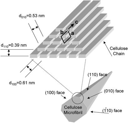
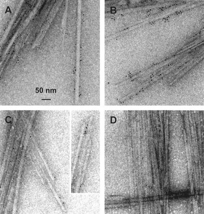
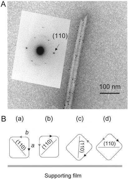
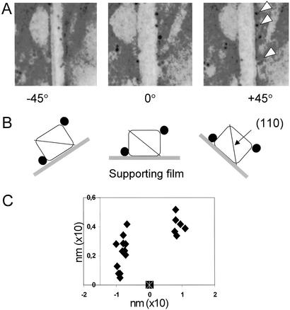
Cellulose binding modules (CBMs) potentiate the action of cellulolytic enzymes on insoluble substrates. Numerous studies have established that three aromatic residues on a CBM surface are needed for binding onto cellulose crystals and that tryptophans contribute to higher binding affinity than tyrosines. However, studies addressing the nature of CBM-cellulose interactions have so far failed to establish the binding site on cellulose crystals targeted by CBMs. In this study, the binding sites of CBMs on Valonia cellulose crystals have been visualized by transmission electron microscopy. Fusion of the CBMs with a modified staphylococcal protein A (ZZ-domain) allowed direct immuno-gold labeling at close proximity of the actual CBM binding site. The transmission electron microscopy images provide unequivocal evidence that the fungal family 1 CBMs as well as the family 3 CBM from Clostridium thermocellum CipA have defined binding sites on two opposite corners of Valonia cellulose crystals. In most samples these corners are worn to display significant area of the hydrophobic (110) plane, which thus constitutes the binding site for these CBMs.


Figures

Figure 1
Figure 1
Schematic presentation of the organization of the cellulose chains in the Iα allomorph of cellulose crystals and the shape of the complete crystal formed. As their unit cell parameters have been determined, Valonia crystals are known to be primarily in the Iα form (12) and the corresponding indexing is thus used throughout this article. The d-spacings characteristic to the different crystalline planes are indicated. The obtuse corner (circled), which exposes the (110) face in worn crystals, is the proposed binding site for the CBMs.
Scheme 1
Scheme 1
Figure 2
Figure 2
Equilibrium adsorption isotherms of different CBM-ZZ fusion proteins: T. reesei CBMCel7A (⧫), T. reesei CBMCel6A (□), T. reesei CBMCel7B (●), N. patriciarum CBMCel6A (+), T. reesei CBMY5W (▵), T. reesei CBMY5W;Y31W (x), isolated T. reesei CBMCel7A (■), and ZZ (○). (A) Adsorption to BMCC (+4°). (B) Adsorption of Cel7A, Cel6A, and mutated CBM-fusion proteins to Valonia cellulose (+4°). (C) Adsorption of ZZ-CBMCel7A and isolated CBMCel7A to BMCC (+22°).
Figure 3
Figure 3
Images obtained by TEM of the labeled CBMs binding to the Valonia cellulose crystals. (A) Cel7A CBM1. (B) Cel7B CBM1. (C) CipA CBM3. (D) Negative control (see text).
Figure 4
Figure 4
(A) Microdiffraction pattern corresponding to the d-spacing of 0.39 nm, which is characteristic of the (110) plane. This plane is parallel to the electron beam in crystal orientation c. (B) Schematic representation of four alternative orientations of the cellulose crystals relative to the underlying carbon support as discussed in the text.
Figure 5
Figure 5
(A) Electron micrographs showing a typical distribution of gold-labeled CBM1Cel7A on an individual Valonia cellulose crystal rotated from the initial 0° position to ±45° along its microfibril axis. The white arrows in A indicate the labels examined and B shows schematically the outcome of the rotation experiment. The black circles represent the labeled CBMs as seen on the cellulose crystals rotated in the three different angles. (C) An example of the 3D mapping of the distribution of the gold particles (filled symbols) recorded in the rotating experiment. Each symbol corresponds to one bound CBM1Cel7A on a single Valonia cellulose crystal. The symbol at the x axis indicates the location of the origin of the coordinate system used (see Materials and Methods for an explanation). (Magnifications: ×250.)

References

    1. Linder M, Teeri T T. J Biotechnol. 1997;57:15–28. - PubMed
    1. Tomme P, Warren R A J, Miller R C, Kilburn D G, Gilkes N R. ACS Symp Ser. 1995;618:143–163.
    1. Reinikainen T, Teleman O, Teeri T T. Proteins. 1995;22:392–403. - PubMed
    1. Kraulis J, Clore G M, Nilges M, Jones T A, Pettersson G, Knowles J, Gronenborn A M. Biochemistry. 1989;28:7241–7257. - PubMed
    1. Mattinen M L, Linder M, Teleman A, Annila A. FEBS Lett. 1997;407:291–296. - PubMed

Publication types

LinkOut - more resources