. 2003 Feb;77(3):1718-26.
doi: 10.1128/jvi.77.3.1718-1726.2003.

The natural host range shift and subsequent evolution of canine parvovirus resulted from virus-specific binding to the canine transferrin receptor

Affiliations

The natural host range shift and subsequent evolution of canine parvovirus resulted from virus-specific binding to the canine transferrin receptor

Karsten Hueffer et al. J Virol. 2003 Feb.

Abstract

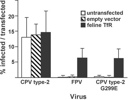
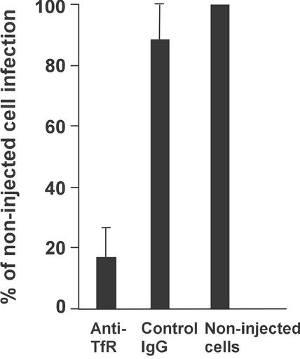
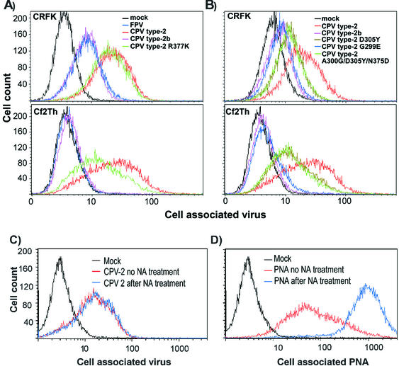
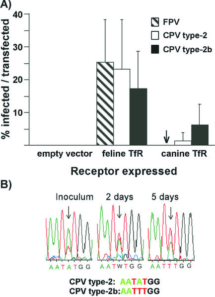
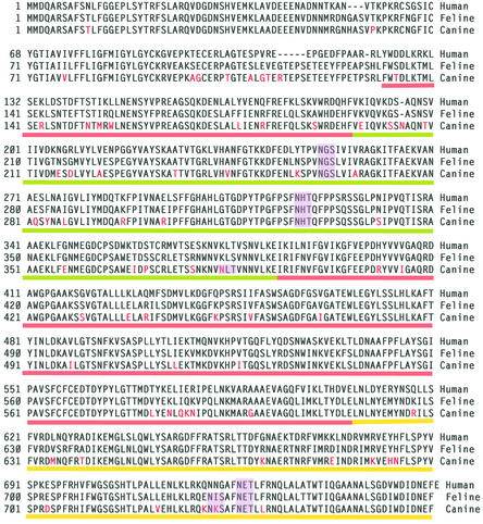
Canine parvovirus (CPV) is a host range variant of a feline virus that acquired the ability to infect dogs through changes in its capsid protein. Canine and feline viruses both use the feline transferrin receptor (TfR) to infect feline cells, and here we show that CPV infects canine cells through its ability to specifically bind the canine TfR. Receptor binding on host cells at 37 degrees C only partially correlated with the host ranges of the viruses, and an intermediate virus strain (CPV type 2) bound to higher levels on cells than did either the feline panleukopenia virus or a later strain of CPV. During the process of adaptation to dogs the later variant strain of CPV gained the ability to more efficiently use the canine TfR for infection and also showed reduced binding to feline and canine cells compared to CPV type 2. Differences on the top and the side of the threefold spike of the capsid surface controlled specific TfR binding and the efficiency of binding to feline and canine cells, and these differences also determined the cell infection properties of the viruses.


Figures

FIG. 1.
FIG. 1.
Expression of the feline TfR in canine Cf2Th cells makes them susceptible to FPV and CPV type 2-G299E infection. Cf2Th cells were transfected with a plasmid expressing the feline TfR or with the empty plasmid vector, incubated for 4 days, and then inoculated with 10 TCID50 per cell of CPV type 2, FPV, or CPV type 2-G299E. After 24 h more incubation, the cells were fixed and infection was detected with an antibody against the NS1 protein. Bars show one standard deviation of the data from three separate experiments.
FIG. 2.
FIG. 2.
Effect of a microinjected antibody against the TfR cytoplasmic tail on CPV infection of Cf2Th cells. Cells were injected with anti-TfR IgG or with a control IgG, inoculated with 2 TCID50 of CPV type 2 per cell, and then incubated for 48 h. Injected cells were identified by staining for the IgG, and infected cells were identified by staining for the viral NS1 protein. The proportion of antibody-injected cells that became infected was compared to the proportion of noninjected cells infected in the same culture. Bars show one standard deviation of the mean for four experiments with the anti-TfR and for two experiments with the control IgG.
FIG. 3.
FIG. 3.
(A) Binding and uptake of FPV (blue), CPV type 2 (red), CPV type 2b (purple), or CPV type 2-R377K (green) capsids incubated with feline CRFK cells or canine Cf2Th cells. Cells were incubated with 20 μg of capsid/ml for 1 h at 37°C and then detached with EDTA, fixed, and permeabilized; the cell-associated virus was quantified with a Cy2-labeled anti-capsid monoclonal antibody, followed by analysis by flow cytometry. (B) Binding and uptake of CPV type 2 (red), CPV type 2b (purple), CPV type 2 D305Y (dark green), CPV type 2 G299E (blue), and CPV type 2 A300G/D305Y/N375D (light green) capsids into CRFK or Cf2Th cells as described above. (C and D) Binding of CPV type 2 capsids to CRFK cells that were either mock treated (red) or incubated with neuraminidase (blue). Cells were incubated with CPV type 2 (C) or fluorescein isothiocyanate-labeled peanut agglutinin (PNA) (D).
FIG. 4.
FIG. 4.
Virus binding to TRVb cells expressing feline or canine TfRs. TRVb cells were transfected with plasmids expressing the feline or canine TfR and then incubated for 4 days. The cells were incubated at 37°C for 1 h with Cy5-labeled canine Tf and with 10 μg of FPV, CPV type 2, CPV type 2b, or CPV type 2-G299E capsids/ml and then washed, suspended by EDTA treatment, fixed, and permeabilized. Capsids were detected with Cy2-labeled antibody. Cell associated Tf is shown on the y axis, and virus capsids are shown on the x axis.
FIG. 5.
FIG. 5.
(A) Virus susceptibility of TRVb cells expressing feline or canine TfRs. Cells transfected with plasmids expressing the feline or canine TfRs were inoculated with CPV type 2, FPV, or CPV type 2b and then incubated for 24 h before incubation for 30 min with Texas Red-labeled canine Tf. After fixation and permeabilization virus infection detected by staining for the viral NS1 protein. The bars represent one standard deviation of the mean of the percentage of Tf-binding cells that became infected in six separate experiments. (B) The replacement of CPV type 2 by CPV type 2a when the two viruses were grown together in TRVb cells expressing the canine TfR for 5 days. The inoculum contained nine times more TCID50 of CPV type 2 than CPV type 2b, which was reflected in the double sequencing profile at position 3046. Samples of virus were collected from the culture inoculum and from the culture at days 2 and 5 after inoculation. The viral DNA was amplified by PCR and sequenced, and the profiles of sequences from nucleotides 3043 to 3049 are shown.
FIG. 6.
FIG. 6.
Aligned sequences of the human, feline, and canine TfRs. The sequence differences that were uniquely seen between the feline and canine TfR sequences are indicated in red. Predicted N-linked glycosylation sites (Asn-X-Ser/Thr) in the different sequences are indicated by shading. The domains of the ectodomain of the receptor determined from the structure of the human TfR are underlined with red (protease-like domain), green (apical domain), and yellow (helical domain) lines.
FIG. 7.
FIG. 7.
(A) Differences between the feline and canine TfR sequences mapped within the structure of the human TfR. One monomer of the ectodomain of the human TfR is shown as a ribbon diagram, while the other monomer is shown as an α-carbon tracing. The side chains of residues in the human TfR model that are unique differences between the feline and canine TfR sequences are indicated in red on one monomer. (B) Models showing the surface of the capsid of CPV (46) and of a dimer of the human TfR ectodomain (17) at the same scale. The monomers of the TfR dimer are shown in red and yellow. (C) The structure of the CPV type 2 capsid in the region that controls host range and canine TfR binding. The polypeptide chains contributed by the four VP2 monomers that make up this region of the capsid are colored differently, and the residues that are discussed in the text as controlling host range or receptor binding are labeled. (D) A road map determined by the method of Rossmann and Palmenberg (32) showing the surface exposure of VP2 residues in one asymmetric unit of the CPV type 2 capsid. The region shown is comprised of several symmetry-related VP2 subunits, and the residues are given, along with the positions in the VP2 sequence. Residues that affect receptor binding or host range and which differ naturally between FPV and CPV strains are outlined in red, whereas residues that were experimentally identified as affecting the feline or canine host ranges of the viruses are indicated in blue.

References

    1. Agbandje, M., R. McKenna, M. G. Rossmann, M. L. Strassheim, and C. R. Parrish. 1993. Structure determination of feline panleukopenia virus empty particles. Proteins 16:155-171. - PubMed
    1. Agbandje-McKenna, M., A. L. Llamas-Saiz, F. Wang, P. Tattersall, and M. G. Rossmann. 1998. Functional implications of the structure of the murine parvovirus, minute virus of mice. Structure 6:1369-1381. - PubMed
    1. Ball-Goodrich, L. J., and P. Tattersall. 1992. Two amino acid substitutions within the capsid are coordinately required for acquisition of fibrotropism by the lymphotropic strain of minute virus of mice. J. Virol. 66:3415-3423. - PMC - PubMed
    1. Barbis, D. P., S.-F. Chang, and C. R. Parrish. 1992. Mutations adjacent to the dimple of canine parvovirus capsid structure affect sialic acid binding. Virology 191:301-308. - PubMed
    1. Bates, G. W., and M. R. Schlabach. 1973. The reaction of ferric salts with transferrin. J. Biol. Chem. 248:3228-3232. - PubMed

Publication types

Substances

LinkOut - more resources