. 2003 Feb;77(3):2010-20.
doi: 10.1128/jvi.77.3.2010-2020.2003.

Importance of basic residues in binding of rous sarcoma virus nucleocapsid to the RNA packaging signal

Affiliations

Importance of basic residues in binding of rous sarcoma virus nucleocapsid to the RNA packaging signal

Eun-gyung Lee et al. J Virol. 2003 Feb.

Erratum in

  • J Virol. 2003 Apr;77(7):4468

Abstract

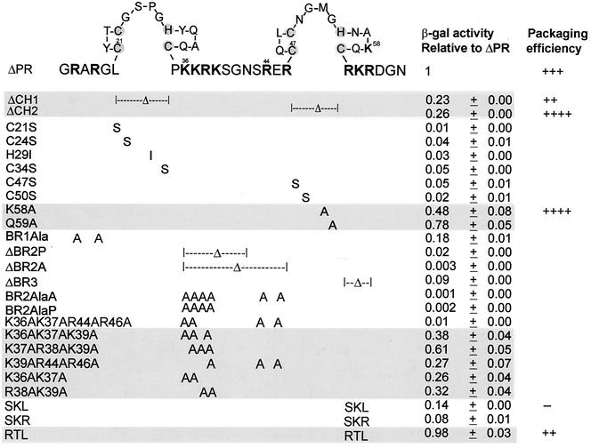
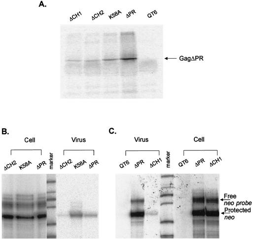
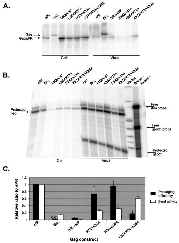
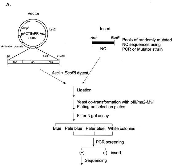
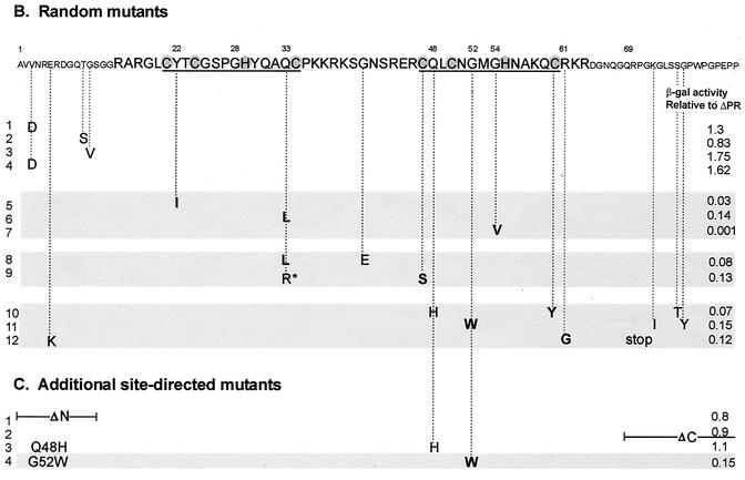
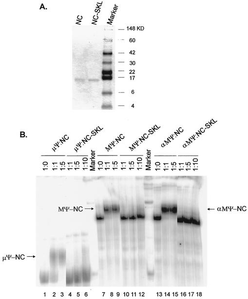
In the context of the Rous sarcoma virus Gag polyprotein, only the nucleocapsid (NC) domain is required to mediate the specificity of genomic RNA packaging. We have previously showed that the Saccharomyces cerevisiae three-hybrid system provides a rapid genetic assay to analyze the RNA and protein components of the avian retroviral RNA-Gag interactions necessary for specific encapsidation. In this study, using both site-directed mutagenesis and in vivo random screening in the yeast three-hybrid binding assay, we have examined the amino acids in NC required for genomic RNA binding. We found that we could delete either of the two Cys-His boxes without greatly abrogating either RNA binding or packaging, although the two Cys-His boxes are likely to be required for efficient viral assembly and release. In contrast, substitutions for the Zn-coordinating residues within the boxes did prevent RNA binding, suggesting changes in the overall conformation of the protein. In the basic region between the two Cys-His boxes, three positively charged residues, as well as basic residues flanking the two boxes, were necessary for both binding and packaging. Our results suggest that the stretches of positively charged residues within NC that need to be in a proper conformation appear to be responsible for selective recognition and binding to the packaging signal (Psi)-containing RNAs.


Figures

FIG. 1.
FIG. 1.
NC site-directed mutations and the resulting β-Gal activities relative to ΔPR. The amino acid sequence of NC is shown using one-letter abbreviations. Amino acid numbering is from the amino terminus of NC. The positively charged residues are indicated in bold type, and the Zn-coordinating residues within the CH boxes are shaded. β-Gal activity relative to that of ΔPR is given as the average ± standard deviation of the values for three to four independent transformants in three assays. The β-Gal activity of cells cotransformed with the RNA hybrid plasmid carrying MΨ sequences and the ΔPR protein hybrid plasmid is about 1,500 U. Mutants with β-Gal activity that was more than 20% of ΔPR are shaded. Packaging efficiencies are summarized from Table 1, Fig. 3 for the SKL mutant, or previous experiments for the RTL mutants (Table 2 in reference 31). Packaging efficiency symbols: ++, the relative packaging efficiency to ΔPR is about 0.8; +++, the relative packaging efficiency to ΔPR is about 1.0; ++++, the relative packaging efficiency to ΔPR is four- to sevenfold more than ΔPR; −, the relative packaging efficiency to ΔPR is about 1% that of ΔPR.
FIG. 2.
FIG. 2.
In vivo packaging assays of the NC mutants. (A) RIPA analysis of pelleted virus-like particles collected from supernatants to quantitate the number of viral particles released to the medium. QT6 cells represent untransfected cells. (B) RPA to measure the amount of neo RNA in cells transfected with either ΔCH2, K58A, or ΔPR mutation or supernatant virions, using an antisense neo RNA as the riboprobe. (C) RPA to measure the effect of the ΔCH1 mutation on the ability of Gag polyprotein to package viral RNA into virions. The amount of neo RNA was measured in transfected cells and virions.
FIG. 2.
FIG. 2.
In vivo packaging assays of the NC mutants. (A) RIPA analysis of pelleted virus-like particles collected from supernatants to quantitate the number of viral particles released to the medium. QT6 cells represent untransfected cells. (B) RPA to measure the amount of neo RNA in cells transfected with either ΔCH2, K58A, or ΔPR mutation or supernatant virions, using an antisense neo RNA as the riboprobe. (C) RPA to measure the effect of the ΔCH1 mutation on the ability of Gag polyprotein to package viral RNA into virions. The amount of neo RNA was measured in transfected cells and virions.
FIG. 3.
FIG. 3.
(A) RIPA to measure the amount of Gag protein in cells transfected with various Gag constructs and to determine the number of virus particles released to the medium. All Gag polyproteins lack the PR domain at the C terminus except SKL, which has a point mutation at the active site in PR. (B) RPA to measure the amount of neo RNA in virions and the amounts of neo RNA and cellular gapdh RNA in transfected cells. The Rnase + lane contains a mixture of the neo probe RNA and gapdh probe RNA treated with RNase cocktail (RNase A and T1). The Rnase − lane was not treated with RNase cocktail. (C) Comparison of the β-Gal activity measured in the yeast three-hybrid binding assays and the packaging efficiency determined in vivo. The packaging efficiency was calculated as follows: the amount of neo RNA in virions (as measured by RPA [shown in panel B]) was normalized to the amount of cellular neo RNA, relative to the level of cellular gapdh RNA (as measured by RPA of whole-cell lysates [shown in panel B]). This calculated neo RNA was then normalized to the number of virions (as measured by RIPA [shown in panel A]). Each experiment was done three times, and the means ± standard deviations (error bars) are shown. Both the β-Gal activity and packaging efficiency were normalized to those for ΔPR. The relative packaging efficiency of the SKL mutant is shown above the bar. The β-Gal activities were summarized from Fig. 1.
FIG. 4.
FIG. 4.
(A) Strategy for in vivo screening of random NC mutants using the yeast three-hybrid system. Pools of randomly mutated NC sequences by using either PCR or a mutator strain were swapped for the wt NC sequences of the pACTIIΔPR-Asc protein hybrid plasmid. The ligated DNAs were used to cotransform yeast cells along with the pIII/MS2-MΨ RNA hybrid plasmid and plated onto plates lacking Ura and Leu to select both plasmids. β-Gal activity of each transformant was qualitatively measured by filter β-Gal assays. Colonies with low β-Gal activities were screened for the NC inserts using PCR. The positive (+) clones were sequenced to locate mutations in the NC sequences. (B) Mutational changes resulting from random mutagenesis and in vivo screening. The amino acid sequence of NC is shown at the top, with the numbering starting from the amino terminus of NC. The two CH boxes are underlined, and the Zn-coordinating residues within the CH boxes are shaded. The mutational changes predicted to significantly lower the β-Gal activity are shown in bold type. The asterisk in line 9 represents a mutational change from glutamine to arginine at residue 33 of NC. A stop mutation at Gln69 (line 12) led to the truncation of NC at the C terminus. The relative β-Gal activities indicated are the averages from three independent assays. The mutant with low β-Gal activity (less than 20% of that of ΔPR) are indicated by shading. (C) Additional site-directed mutants. ΔN has residues 1 to 13 of NC deleted; ΔC lacks all of the amino acids following position 68. The G52W mutant which had β-Gal activity of less than 0.2 is shaded.
FIG. 4.
FIG. 4.
(A) Strategy for in vivo screening of random NC mutants using the yeast three-hybrid system. Pools of randomly mutated NC sequences by using either PCR or a mutator strain were swapped for the wt NC sequences of the pACTIIΔPR-Asc protein hybrid plasmid. The ligated DNAs were used to cotransform yeast cells along with the pIII/MS2-MΨ RNA hybrid plasmid and plated onto plates lacking Ura and Leu to select both plasmids. β-Gal activity of each transformant was qualitatively measured by filter β-Gal assays. Colonies with low β-Gal activities were screened for the NC inserts using PCR. The positive (+) clones were sequenced to locate mutations in the NC sequences. (B) Mutational changes resulting from random mutagenesis and in vivo screening. The amino acid sequence of NC is shown at the top, with the numbering starting from the amino terminus of NC. The two CH boxes are underlined, and the Zn-coordinating residues within the CH boxes are shaded. The mutational changes predicted to significantly lower the β-Gal activity are shown in bold type. The asterisk in line 9 represents a mutational change from glutamine to arginine at residue 33 of NC. A stop mutation at Gln69 (line 12) led to the truncation of NC at the C terminus. The relative β-Gal activities indicated are the averages from three independent assays. The mutant with low β-Gal activity (less than 20% of that of ΔPR) are indicated by shading. (C) Additional site-directed mutants. ΔN has residues 1 to 13 of NC deleted; ΔC lacks all of the amino acids following position 68. The G52W mutant which had β-Gal activity of less than 0.2 is shaded.
FIG. 5.
FIG. 5.
Binding of Ψ RNA to the NC proteins. (A) Purification of NC proteins. wt NC and a mutant version of NC (NC-SKL) were expressed, purified by fast-performance liquid chromatography, electrophoresed on a SDS-10% polyacrylamide gel, and stained with Coomassie brilliant blue. The positions of molecular mass markers (in kilodaltons) are shown to the right of the gel. (B) In vitro-transcribed RNA was not mixed or mixed with NC or NC-SKL proteins at the indicated molar ratios. The band shifts are indicated by arrows. μΨ, an 82-nt minimal packaging sequence; MΨ, a 160-nt packaging sequence; αMΨ, antisense MΨ RNA.

References

    1. Aronoff, R., and M. L. Linial. 1991. Specificity of retroviral RNA packaging. J. Virol. 65:71-80. - PMC - PubMed
    1. Banks, J. D., and M. L. Linial. 2000. Secondary structure analysis of a minimal avian leukosis-sarcoma virus packaging signal. J. Virol. 74:456-464. - PMC - PubMed
    1. Banks, J. D., A. Yeo, K. Green, F. Cepeda, and M. L. Linial. 1998. A minimal avian retroviral packaging sequence has a complex structure. J. Virol. 72:6190-6194. - PMC - PubMed
    1. Bartel, P. L., J. A. Roecklein, D. SenGupta, and S. Fields. 1996. A protein linkage map of Escherichia coli bacteriophage T7. Nat. Genet. 12:72-77. - PubMed
    1. Bennett, R. P., T. D. Nelle, and J. W. Wills. 1993. Functional chimeras of the Rous sarcoma virus and human immunodeficiency virus Gag proteins. J. Virol. 67:6487-6498. - PMC - PubMed

Publication types

LinkOut - more resources