. 2003 Feb;77(3):2134-46.
doi: 10.1128/jvi.77.3.2134-2146.2003.

Establishment of B-cell lymphoma cell lines persistently infected with hepatitis C virus in vivo and in vitro: the apoptotic effects of virus infection

Affiliations

Establishment of B-cell lymphoma cell lines persistently infected with hepatitis C virus in vivo and in vitro: the apoptotic effects of virus infection

Vicky M-H Sung et al. J Virol. 2003 Feb.

Abstract

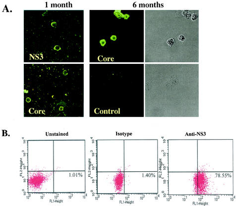
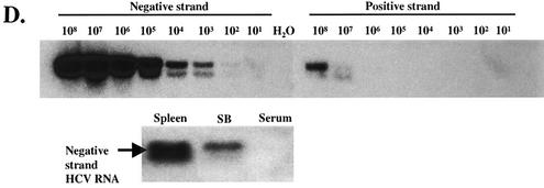
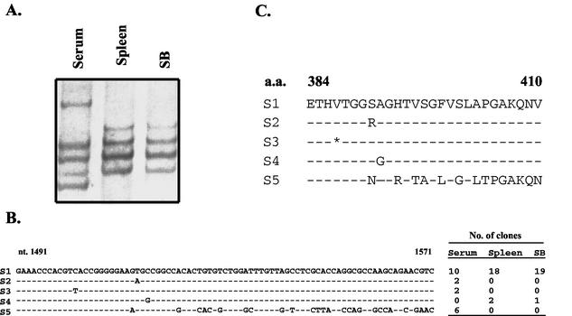
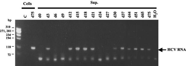
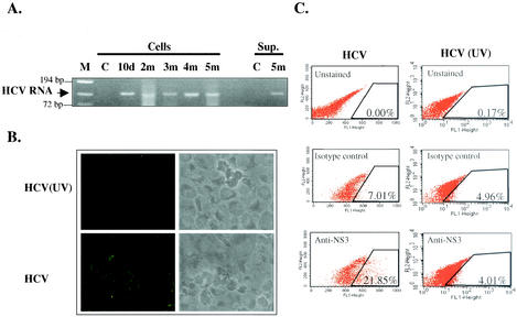
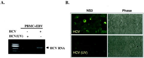
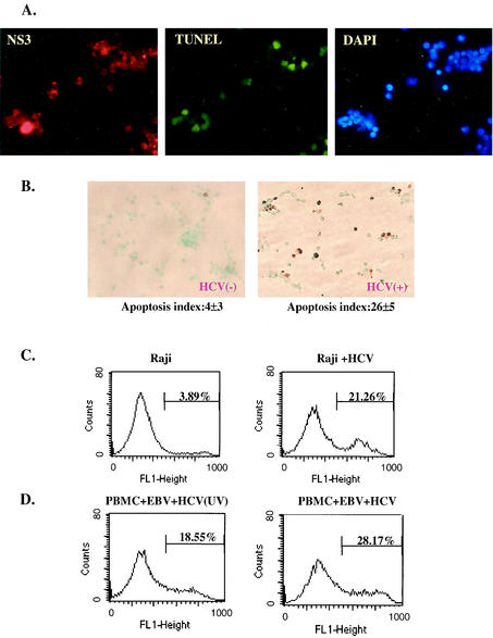
Hepatitis C virus (HCV) is a major cause of chronic hepatitis, liver cirrhosis, and hepatocellular carcinoma. Studies of HCV replication and pathogenesis have so far been hampered by the lack of an efficient tissue culture system for propagating HCV in vitro. Although HCV is primarily a hepatotropic virus, an increasing body of evidence suggests that HCV also replicates in extrahepatic tissues in natural infection. In this study, we established a B-cell line (SB) from an HCV-infected non-Hodgkin's B-cell lymphoma. HCV RNA and proteins were detectable by RNase protection assay and immunoblotting. The cell line continuously produces infectious HCV virions in culture. The virus particles produced from the culture had a buoyant density of 1.13 to 1.15 g/ml in sucrose and could infect primary human hepatocytes, peripheral blood mononuclear cells (PBMCs), and an established B-cell line (Raji cells) in vitro. The virus from SB cells belongs to genotype 2b. Single-stranded conformational polymorphism and sequence analysis of the viral RNA quasispecies indicated that the virus present in SB cells most likely originated from the patient's spleen and had an HCV RNA quasispecies pattern distinct from that in the serum. The virus production from the infected primary hepatocytes showed cyclic variations. In addition, we have succeeded in establishing several Epstein-Barr virus-immortalized B-cell lines from PBMCs of HCV-positive patients. Two of these cell lines are positive for HCV RNA as detected by reverse transcriptase PCR and for the nonstructural protein NS3 by immunofluorescence staining. These observations unequivocally establish that HCV infects B cells in vivo and in vitro. HCV-infected cell lines show significantly enhanced apoptosis. These B-cell lines provide a reproducible cell culture system for studying the complete replication cycle and biology of HCV infections.


Figures

FIG. 1.
FIG. 1.
SB cells are persistently infected by HCV. (A) Detection of HCV protein expression by immunofluorescence staining in SB cells at different time points of culture. Uninfected Raji cells were used as a control. Phase contrast images of SB cells (upper) and uninfected Raji cells (lower) are shown on the right. (B) Detection of intracellular NS3 protein expression in SB cells by flow cytometry. Unstained SB cells served as the “no-antibody” control; isotype control antibody served as the background staining control. (C) Detection of positive-strand HCV RNA in SB cells by RPA. The expected size of the protected band from HCV RNA in SB cells is 123 bp. Different amounts of in vitro-transcribed sense and antisense HCV RNA (IVT-S and IVT-AS) were used for quantitation. The AS probe was used for detecting positive-strand RNA, and the S probe was used for detecting negative-strand RNA. Because of the presence of vector sequences in both the probes and the in vitro-transcribed RNA, the protected sizes for IVT-S and IVT-AS are 215 and 233 bp, respectively. HCV(+), SB RNA hybridized to the AS probe; HCV(-), same RNA hybridized to the S probe. (D) Detection of negative-strand HCV RNA by strand-specific RT-PCR and Southern blotting. The top blot shows the validation of the strand specificity of the assay. Serial dilutions of both positive- and negative-strand in vitro-transcribed HCV RNAs from 108 molecules to 1 molecule were subjected to negative-strand-specific RT-PCR using the sense primer for reverse transcription. The bottom blot shows that negative-strand HCV RNA was detected in the spleen and SB cells but not in the patient's serum.
FIG. 1.
FIG. 1.
SB cells are persistently infected by HCV. (A) Detection of HCV protein expression by immunofluorescence staining in SB cells at different time points of culture. Uninfected Raji cells were used as a control. Phase contrast images of SB cells (upper) and uninfected Raji cells (lower) are shown on the right. (B) Detection of intracellular NS3 protein expression in SB cells by flow cytometry. Unstained SB cells served as the “no-antibody” control; isotype control antibody served as the background staining control. (C) Detection of positive-strand HCV RNA in SB cells by RPA. The expected size of the protected band from HCV RNA in SB cells is 123 bp. Different amounts of in vitro-transcribed sense and antisense HCV RNA (IVT-S and IVT-AS) were used for quantitation. The AS probe was used for detecting positive-strand RNA, and the S probe was used for detecting negative-strand RNA. Because of the presence of vector sequences in both the probes and the in vitro-transcribed RNA, the protected sizes for IVT-S and IVT-AS are 215 and 233 bp, respectively. HCV(+), SB RNA hybridized to the AS probe; HCV(-), same RNA hybridized to the S probe. (D) Detection of negative-strand HCV RNA by strand-specific RT-PCR and Southern blotting. The top blot shows the validation of the strand specificity of the assay. Serial dilutions of both positive- and negative-strand in vitro-transcribed HCV RNAs from 108 molecules to 1 molecule were subjected to negative-strand-specific RT-PCR using the sense primer for reverse transcription. The bottom blot shows that negative-strand HCV RNA was detected in the spleen and SB cells but not in the patient's serum.
FIG. 2.
FIG. 2.
HCV quasispecies of the SB cell. (A) SSCP analysis of HCV quasispecies from the patient. (B) Nucleotide sequences of the HVR (nucleotides [nt] 1491 to 1571) of HCV RNA from SB cells (1 month old) and spleen and serum of the same patient. HVR1 was amplified by nested RT-PCR, and the PCR products were cloned into the pCRII-TOPO vector (Invitrogen). Twenty independent clones from each sample were sequenced from both ends. (C) Amino acid sequences of the HVR (amino acids [a.a.] 384 to 410) deduced from the sequences in panel B. The dashes represent nucleotides or amino acids identical to those of S1. The asterisk represents a silent mutation.
FIG. 3.
FIG. 3.
Establishment of B-cell lines from PBMCs of HCV-infected patients by EBV immortalization. Shown are indirect immunofluorescence staining of HCV NS3 protein on EBV-immortalized B cells from patients 011A (A) and 016A (B) after 2 months in culture (top) and detection of HCV RNA by RT-PCR after 1 month in culture (bottom). The 5′ UTR was used for RT-PCR. PCR without an RT reaction was used as a negative control for cDNA contamination.
FIG. 4.
FIG. 4.
SB cells continuously produce HCV particles. (A) On the left is shown the detection of HCV RNA by RT-PCR from SB cells and culture supernatant. Uninfected Raji cells were used as a negative control. On the right is shown the quantitation by real-time RT-PCR of HCV RNA in the SB cell culture supernatant during 5 days of culture after medium change. One hundred million SB cells were cultured in 10 ml of medium, and 1 ml was harvested for RT-PCR every day. (B) Western blotting of HCV NS3 (top) and core protein (bottom) in the cell lysate (Cells) and culture supernatant (Sup.). The asterisks indicate HCV core or NS3 protein. (C) Equilibrium sucrose gradient sedimentation of the SB cell culture supernatant. The HCV RNA in each fraction was measured by real-time RT-PCR.
FIG. 5.
FIG. 5.
HCV produced from SB cells can infect primary human hepatocytes. Virus produced from primary human hepatocytes infected with SB cell culture supernatant (Sup.) was analyzed by RT-PCR of HCV RNA. Uninfected primary human hepatocytes (Cells) were used as a control (C). Cellular RNA on day 75 (d75) was used as the positive control.
FIG. 6.
FIG. 6.
Infection of Raji cells by HCV from SB cells. (A) Continuous detection of HCV RNA in HCV-infected Raji cells. Raji cells were inoculated with SB culture supernatant (Sup.). Cells or culture supernatant were harvested at various times (d, day; m, month) and used for RT-PCR analysis. C, uninfected Raji cells; M, molecular size marker. (B) Detection of HCV NS3 protein by indirect immunofluorescence staining in HCV-infected Raji cells. Raji cells infected with UV-irradiated SB supernatant [HCV (UV)] were used as a control. (C) Detection of intracellular NS3 protein expression in HCV-infected Raji cells (left) and Raji cells infected with UV-irradiated HCV (right) by flow cytometry. (D) Single-cell cloning of HCV-infected Raji cells. HCV-infected Raji cells were diluted into single cells and grown in a 96-well U-bottom plate. After 2 to 3 weeks, 81 clones were obtained and analyzed by RT-PCR to detect HCV RNA. Sixty-four clones were positive for HCV RNA. A through G designate different groups of cell clones processed for RT-PCR.
FIG. 6.
FIG. 6.
Infection of Raji cells by HCV from SB cells. (A) Continuous detection of HCV RNA in HCV-infected Raji cells. Raji cells were inoculated with SB culture supernatant (Sup.). Cells or culture supernatant were harvested at various times (d, day; m, month) and used for RT-PCR analysis. C, uninfected Raji cells; M, molecular size marker. (B) Detection of HCV NS3 protein by indirect immunofluorescence staining in HCV-infected Raji cells. Raji cells infected with UV-irradiated SB supernatant [HCV (UV)] were used as a control. (C) Detection of intracellular NS3 protein expression in HCV-infected Raji cells (left) and Raji cells infected with UV-irradiated HCV (right) by flow cytometry. (D) Single-cell cloning of HCV-infected Raji cells. HCV-infected Raji cells were diluted into single cells and grown in a 96-well U-bottom plate. After 2 to 3 weeks, 81 clones were obtained and analyzed by RT-PCR to detect HCV RNA. Sixty-four clones were positive for HCV RNA. A through G designate different groups of cell clones processed for RT-PCR.
FIG. 7.
FIG. 7.
In vitro Infection of normal PBMCs with HCV from SB cells. (A) Fresh PBMCs were coinfected with EBV and SB supernatant (HCV) or UV-irradiated SB supernatant [HCV (UV)]. Cells and culture media from 1-month-old culture were harvested, and HCV RNA was detected by RT-PCR. +, present; −, absent. (B) The same cells described in the legend to panel A were used for detection of HCV NS3 protein by immunofluorescence staining 3 months postinfection. Phase, phase-contrast image.
FIG. 8.
FIG. 8.
HCV infection causes apoptosis in HCV-infected B cells. (A) SB cells were costained with anti-HCV NS3 antibody, fluorescence-labeled deoxynucleoside triphosphate for TUNEL assay, and DAPI for visualization of all cells. Most of the TUNEL-positive cells were also positive for NS3. (B) Detection of apoptotic cells by TUNEL assay in uninfected Raji cells (left) and HCV-infected Raji cells (right). Apoptotic cells were stained brown. The percentage of apoptotic cells was determined as the average of four fields under the microscope. This experiment was repeated five times. The range of percentages of apoptosis was 1 to 5% for Raji cells and 21 to 29% for HCV-infected Raji cells. (C) Analysis of apoptotic cells by fluorescence-activated cell sorter in HCV-infected (Raji + HCV) and uninfected Raji cells by annexin V-binding assay. (D) Analysis of apoptotic-cell populations in PBMCs coinfected with EBV and SB culture supernatant (HCV) or UV-irradiated SB supernatant [HCV (UV)]. The assays were done on the cells 2 months postinfection.

References

    1. Andre, P., F. Komurian-Pradel, S. Deforges, M. Perret, J. L. Berland, M. Sodoyer, S. Pol, C. Brechot, G. Paranhos-Baccala, and V. Lotteau. 2002. Characterization of low- and very-low-density hepatitis C virus RNA-containing particles. J. Virol. 76:6919-6928. - PMC - PubMed
    1. Blight, K. J., A. A. Kolykhalov, and C. M. Rice. 2000. Efficient initiation of HCV RNA replication in cell culture. Science 290:1972-1974. - PubMed
    1. Bouvier-Alias, M., K. Patel, H. Dahari, S. Beaucourt, P. Larderie, L. Blatt, C. Hezode, G. Picchio, D. Dhumeaux, A. U. Neumann, J. G. McHutchison, and J.-M. Pawlotsky. 2002. Clinical utility of total HCV core antigen quantification: a new indirect marker of HCV replication. Hepatology 36:211-218. - PubMed
    1. Cerny, A., and F. V. Chisari. 1999. Pathogenesis of chronic hepatitis C: immunological features of hepatic injury and viral persistence. Hepatology 30:595-601. - PubMed
    1. Chomczynski, P., and N. Sacchi. 1987. Single-step method of RNA isolation by acid guanidinium thiocyanate-phenol-chloroform extraction. Anal. Biochem. 162:156-159. - PubMed

Publication types

Substances

LinkOut - more resources