. 2003 Jan 15;546(Pt 2):483-90.
doi: 10.1113/jphysiol.2002.031773.

Parallel decrease in omega-conotoxin-sensitive transmission and dopamine-induced inhibition at the striatal synapse of developing rats

Affiliations

Parallel decrease in omega-conotoxin-sensitive transmission and dopamine-induced inhibition at the striatal synapse of developing rats

Toshihiko Momiyama. J Physiol. .

Abstract

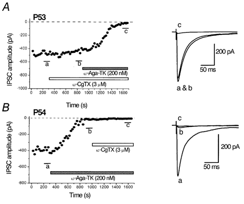
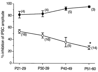
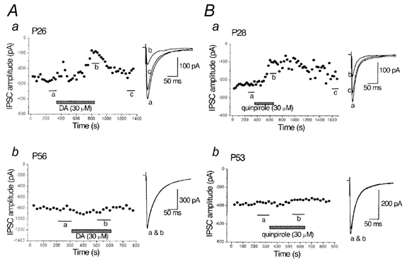
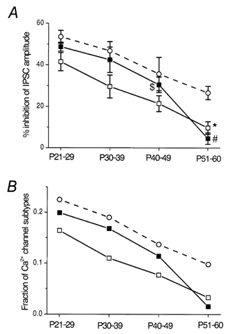
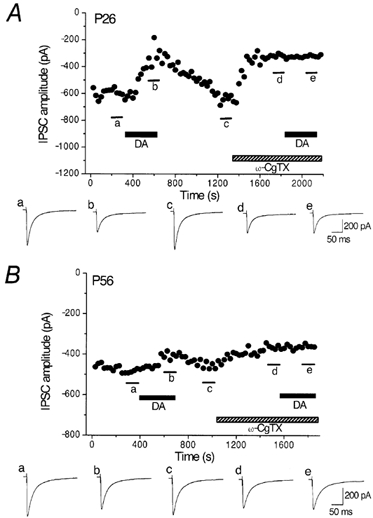
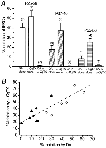
Whole-cell patch-clamp recordings of GABAergic IPSCs were made from cholinergic interneurones in slices of striatum from developing rats aged 21-60 days postnatal. In addition, the Ca(2+) channel subtypes involved in synaptic transmission, as well as dopamine (DA)-induced presynaptic inhibition, were investigated pharmacologically with development by bath application of Ca(2+) channel blockers and DA receptor agonists. The IPSC amplitude was reduced by omega-conotoxin GVIA (omega-CgTX) or omega-agatoxin TK (omega-Aga-TK) across the whole age range, suggesting that multiple types of Ca(2+) channels mediate transmission of the synapse. The IPSC fraction reduced by omega-CgTX significantly decreased, whereas that reduced by omega-Aga-TK remained unchanged with development. DA or quinpirole, a D(2)-like receptor agonist, presynaptically reduced the IPSC amplitude throughout development. The DA-induced inhibition decreased with age in parallel with the decrease in N-type Ca(2+) channels. DA showed no further inhibition of IPSCs after the inhibitory effect of omega-CgTX had reached steady state throughout development. These results demonstrate that there is a functional link between presynaptic N-type Ca(2+) channels and D(2)-like DA receptors at inhibitory synapses in the striatum. They also demonstrate that the suppression of GABAergic transmission by D(2)-like receptors is mediated by modulation of N-type Ca(2+) channels and decreases in parallel with the developmental decline in the contribution of N-type Ca(2+) channels to exocytosis.


Figures

Figure 1
Figure 1. Effects of ω-CgTX and ω-Aga-TK on the GABAergic IPSCs in striatal cholinergic interneurones of adult rats
A, time course of the effects of ω-CgTX (3 μm) and subsequent application of ω-Aga-TK (200 nm) on the amplitude of IPSCs evoked in striatal cholinergic interneurones of a P53 rat. B, time course of the effects of ω-Aga-TK (200 nm) and subsequent application of ω-CgTX (3 μm) on the amplitude of IPSCs in a P54 rat. IPSCs were evoked at 0.2 Hz at −60 mV. Each point represents the mean amplitude of five consecutive responses. Blockers were applied during the indicated periods. Superimposed traces on the right are averages of 20 consecutive IPSCs during the indicated periods.
Figure 2
Figure 2. Summary of the developmental changes in the effects of ω-CgTX and ω-Aga-TK on the IPSCs in striatal cholinergic interneurones
The ω-CgTX-sensitive fraction (○) and ω-Aga-TK-sensitive fraction (•) of the IPSCs are plotted at different postnatal ages. Each point shows the mean ± s.e.m. derived from 3-16 cells, as indicated in parentheses. *P < 0.05 compared with the value of P21-29 or P30-39.
Figure 3
Figure 3. Developmental decline in the inhibitory effects of dopamine (DA) and quinpirole on the IPSCs in striatal cholinergic interneurones
A, time course of the inhibitory effect of DA (30 μm) on the evoked IPSCs in striatal cholinergic interneurones obtained from P26 rats (a) and lack of DA-induced inhibition in a P56 rat (b). B, time course of the inhibitory effect of quinpirole (30 μm) on the IPSCs in a P28 rat (a) and lack of effect in a P53 rat (b). IPSCs were evoked at 0.2 Hz at −60 mV. Each point represents the mean amplitude of five consecutive responses. DA or quinpirole was applied during the indicated periods. Traces to the right show averaged IPSCs of 20 consecutive responses during the indicated periods.
Figure 4
Figure 4. Summarized plots showing the developmental decreases in DA- and quinpirole-induced inhibition of the IPSCs
A, summarized effects of DA (30 μm, □) and quinpirole (30 μm, ▪) on the evoked IPSCs at different postnatal ages, compared with the effect of ω-CgTX (○, derived from Fig. 2). Each point shows the mean ± s.e.m. derived from 3-16 cells. *P < 0.05 compared with the value of P21-29 or P30-39. #P < 0.05 compared with the value of P21-29, P30-39 or P40-49. $P < 0.05 compared with the value of P21-29. B, developmental declines in the estimated fractions of Ca2+ channels involved in the IPSC inhibition by DA (□) or quinpirole (▪), compared with the change in the fraction of N-type channels (○). The fractions were calculated assuming a third-power relation between presynaptic Ca2+ concentration and postsynaptic response amplitude.
Figure 5
Figure 5. Occlusion of DA-induced inhibition by ω-CgTX in young and adult rats
Top panels in A and B, effects of DA (30 μm) on the evoked IPSCs before and after application of ω-CgTX (3 μm) at P26 (A) and P56 (B). IPSCs were evoked at 0.2 Hz. Vh, −60 mV. Each point represents the mean amplitude of five consecutive IPSCs. DA and ω-CgTX were applied in the bath during the indicated periods. ω-CgTX was applied after IPSCs had recovered from the inhibition by an initial application of DA. DA was applied again after ω-CgTX-induced suppression had reached steady state. Bottom panels in A and B, averaged traces of 20 consecutive IPSCs during the indicated periods. Note the small effect of DA and ω-CgTX at P56.
Figure 6
Figure 6. Parallel decrease in DA- and ω-CgTX-induced inhibition with age
A, summarized histograms showing the inhibitory effects of DA (30 μm), ω-CgTX (3 μm) and DA in the presence of ω-CgTX examined in the same neurones at three different age ranges of rats. Note that, throughout the developmental stages, DA no longer inhibited the IPSCs after the effect of ω-CgTX had reached steady state (1.1 ± 0.42 % at P25-28, 0.67 ± 0.39 % at P37-40 and 1.6 ± 0.54 % at P55-56). Numbers of cells are indicated in parentheses. B, correlation between DA- and ω-CgTX-induced inhibition of the IPSCs examined in the same neurones. Each point represents one neurone summarized in A (○, P25-28; •, P37-40; ▴, P55-56). The dashed line was drawn by the least-squares method (r = 0.83, P < 0.001).

References

    1. Aosaki T, Kiuchi K, Kawaguchi Y. Dopamine D1-like receptor activation excites rat striatal large aspiny neurons in vitro. J Neurosci. 1998;18:5180–5190. - PMC - PubMed
    1. Barnes-Davies M, Owens S, Forsythe ID. Calcium channels triggering transmitter release in the rat medial superior olive. Hear Res. 2001;162:134–145. - PubMed
    1. Bolam P, Wainer BH, Smith AD. Characterization of cholinergic neurons in the rat neostriatum. A combination of choline acetyltransferase immunocytochemistry, Golgi-impregnation and electron microscopy. Neurosci. 1984;12:711–718. - PubMed
    1. Broaddus WC, Bennett JP., Jr Postnatal development of striatal dopamine function. I. An examination of D1 and D2 receptors, adenylate cyclase regulation and presynaptic dopamine markers. Brain Res Mol Brain Res. 1990;52:265–271. - PubMed
    1. Calabresi P, Centonze D, Gubellini P, Pisani A, Bernardi G. Acetylcholine-mediated modulation of striatal function. Trends Neurosci. 2000;23:120–126. - PubMed

Publication types

MeSH terms

LinkOut - more resources