. 2003 Jan 20;160(2):245-53.
doi: 10.1083/jcb.200208109. Epub 2003 Jan 13.

Binding of an ankyrin-1 isoform to obscurin suggests a molecular link between the sarcoplasmic reticulum and myofibrils in striated muscles

Affiliations

Binding of an ankyrin-1 isoform to obscurin suggests a molecular link between the sarcoplasmic reticulum and myofibrils in striated muscles

Paola Bagnato et al. J Cell Biol. .

Abstract

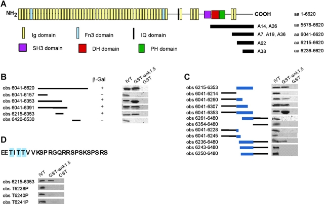
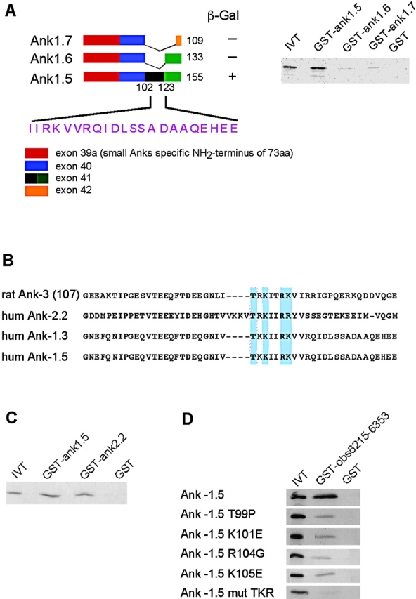
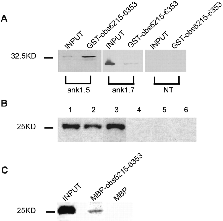
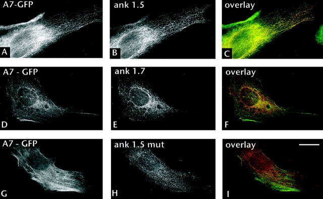
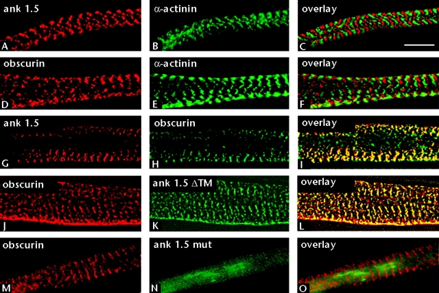
Assembly of specialized membrane domains, both of the plasma membrane and of the ER, is necessary for the physiological activity of striated muscle cells. The mechanisms that mediate the structural organization of the sarcoplasmic reticulum with respect to the myofibrils are, however, not known. We report here that ank1.5, a small splice variant of the ank1 gene localized on the sarcoplasmic reticulum membrane, is capable of interacting with a sequence of 25 aa located at the COOH terminus of obscurin. Obscurin is a giant sarcomeric protein of approximately 800 kD that binds to titin and has been proposed to mediate interactions between myofibrils and other cellular structures. The binding sites and the critical aa required in the interaction between ank1.5 and obscurin were characterized using the yeast two-hybrid system, in in vitro pull-down assays and in experiments in heterologous cells. In differentiated skeletal muscle cells, a transfected myc-tagged ank1.5 was found to be selectively restricted near the M line region where it colocalized with endogenous obscurin. The M line localization of ank1.5 required a functional obscurin-binding site, because mutations of this domain resulted in a diffused distribution of the mutant ank1.5 protein in skeletal muscle cells. The interaction between ank1.5 and obscurin represents the first direct evidence of two proteins that may provide a direct link between the sarcoplasmic reticulum and myofibrils. In keeping with the proposed role of obscurin in mediating an interaction with ankyrins and sarcoplasmic reticulum, we have also found that a sequence with homology to the obscurin-binding site of ank1.5 is present in the ank2.2 isoform, which in striated muscles has been also shown to associate with the sarcoplasmic reticulum. Accordingly, a peptide containing the COOH terminus of ank2.2 fused with GST was found to bind to obscurin. Based on reported evidence showing that the COOH terminus of ank2.2 is necessary for the localization of ryanodine receptors and InsP3 receptors in the sarcoplasmic reticulum, we propose that obscurin, through multiple interactions with ank1.5 and ank2.2 isoforms, may assemble a large protein complex that, in addition to a structural function, may play a role in the organization of specific subdomains in the sarcoplasmic reticulum.


Figures

Figure 1.
Figure 1.
Association of ank1.5 with the COOH-terminal region of obscurin. (A) Two-hybrid screening with the cytosolic tail of ank1.5 (aa 22–155) as “bait” identified seven positive clones, corresponding to the COOH terminus of the obscurin protein. (B) Fragments of the obscurin clone A7 were tested for the ability to interact with the bait ank1.5 in the two-hybrid system and with a GST–ank1.5 fusion protein in pull-down experiments. Both experiments demonstrated that the region of obscurin between aa 6236 and 6353 is capable to interact with ank1.5. +, detectable activity; −, no detectable activity. (C) A second series of obscurin fragments was prepared to further restrict the sequence responsible for binding with ank1.5 to 25 aa of obscurin (aa 6236–6260). (D) Mutagenesis of the aa in the COOH terminus of obscurin (aa 6236–6260) capable of mediating binding to ank1.5 was performed and mutant proteins were tested against GST–ank1.5 in in vitro binding assays.
Figure 2.
Figure 2.
The site in ank1.5 able to interact with obscurin is located in a sequence highly conserved among ankyrins. (A) A schematic representation of the structure of the small muscle–specific ankyrin isoforms is presented: a color code identifies the different exons that, by undergoing alternative splicing, generate the different ank1 isoforms. The cytosolic sequences of ank1.5, ank1.6, and ank1.7 were used as baits in two-hybrid system experiments against the obscurin subclone A7. In parallel, in vitro–translated obscurin subclone A7 was allowed to interact with GST–ank1.5, GST–ank1.6, and GST–ank1.7. Both two-hybrid and in vitro binding assays demonstrated that only ank1.5 is capable of binding the obscurin subclone A7. +, detectable activity; −, no detectable activity. (B) The alignment of COOH-terminal sequences of a skeletal muscle–specific ank3 isoform (AnkG107), ank2.2, ank1.3, and ank1.5. Conserved aa residues are in bold. (C) In vitro binding of a GST fusion protein containing the COOH-terminal region of ank2.2 (aa 1758–1872) against in vitro–transcribed and –translated obscurin clone A7. (D) Site-directed mutagenesis of aa 97–123 of ank1.5 was performed and the in vitro–transcribed and –translated mutant proteins were used in binding experiments with the GST–Obs6215–6353 fusion protein. Mutagenesis of any of four aa residues (T99, K101, R104, and K105) of ank1.5 strongly reduced the ability to bind to obscurin.
Figure 3.
Figure 3.
Exogenous and endogenous ank1.5 interacts with the COOH-terminal region of obscurin. (A) Microsomes from HEK293 cells transfected with myc-tagged full-length cDNAs of ank1.5 and ank1.7 were used in interaction studies with the GST–Obs6215–6353 fusion protein. (B) ank1.5 is present in the sarcoplasmic reticulum of skeletal muscle. ank1.5 reactivity was detected in the total microsomal fraction (lane 1), in sarcoplasmic reticulum fractions enriched in longitudinal tubules (lane 2), and in terminal cisternae (lane 3). No signal was detected by a preimmune sera when tested against the same preparations of the total microsomal fraction (lane 4), longitudinal tubules (lane 5), and terminal cisternae (lane 5). (C) Microsomes from mouse skeletal muscles were used as a source of endogenous ank1.5 to perform pull-down experiments against the MBP–Obs6215–6353 fusion protein.
Figure 4.
Figure 4.
Effect of ank1.5 on the localization of obscurin in NIH 3T3 cells. NIH 3T3 cells were transfected with the obscurin subclone A7 joined in frame with GFP, the full-length cDNA of ank1.5, ank1.7, and mutant ank1.5 cloned in pcDNA3 vectors. Cells were decorated with anti-myc monoclonal antibodies and detected by a Cy3-conjugated anti–mouse antibody (red). Panels show representative sections from the top to bottom of the cells at ∼0.25-μm intervals. Bar, 5 μm.
Figure 5.
Figure 5.
Localization of ank1.5 and obscurin in differentiating skeletal muscle cells. Differentiated skeletal muscle cells were stained with rabbit antibodies against the myc epitope to recognize the myc-tagged ank1.5 protein (A) and with monoclonal antibodies against α-actinin, which stain the Z line (B). The alternating pattern of ank1.5 and α-actinin is compatible with the localization of ank1.5 near or at the M line. Cells were stained with rabbit antibodies against the COOH-terminal region of obscurin (D) and with monoclonal antibodies against α-actinin (E). Again, the obscurin staining is compatible with localization near or at the M line. Staining of the cells with antibodies against ank1.5 (G) and against obscurin (H) indicates that the two proteins colocalize. Soluble ank1.5 (ank1.5 ΔTM) missing the transmembrane domain (K) is still able to colocalize with obscurin at the M line (L). A myc-tagged mutant ank1.5, unable to bind to obscurin, presents a diffuse signal (N) distinct from that of obscurin (M). Bar, 5 μm.

References

    1. Bang, M.L., T. Centner, F. Fornoff, A.J. Geach, M. Gotthardt, M. McNabb, C.C. Witt, D. Labeit, C.C. Gregorio, H. Granzier, and S. Labeit. 2001. The complete gene sequence of titin, expression of an unusual approximately 700-kDa titin isoform, and its interaction with obscurin identify a novel Z-line to I-band linking system. Circ. Res. 89:1065–1072. - PubMed
    1. Baumann, O., and B. Walz. 2001. Endoplasmic reticulum of animal cells and its organization into structural and functional domains. Int. Rev. Cytol. 205:149–214. - PubMed
    1. Beck, K.A., J.A. Buchanan, and W.J. Nelson. 1997. Golgi membrane skeleton: identification, localization and oligomerization of a 195 kDa ankyrin isoform associated with the Golgi complex. J. Cell Sci. 110:1239–1249. - PubMed
    1. Bellin, R.B., T.W. Huiatt, D.R. Critchley, and R.M. Robson. 2001. Synemin may function to directly link muscle cell intermediate filaments to both myofibrillar Z-lines and costameres. J. Biol. Chem. 276:32330–32337. - PubMed
    1. Bennett, V., and A.J. Baines. 2001. Spectrin and ankyrin-based pathways: metazoan invenctions for integrating cells into tissues. Physiol. Rev. 81:1353–1392. - PubMed

Publication types

MeSH terms