. 2003 Jan 15;31(2):532-50.
doi: 10.1093/nar/gkg161.

Structural classification of zinc fingers: survey and summary

Affiliations

Structural classification of zinc fingers: survey and summary

S Sri Krishna et al. Nucleic Acids Res. .

Abstract

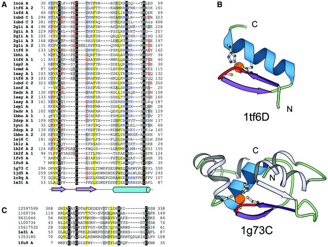
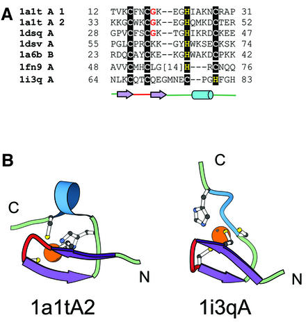
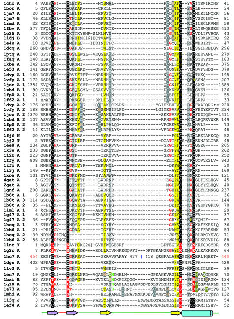
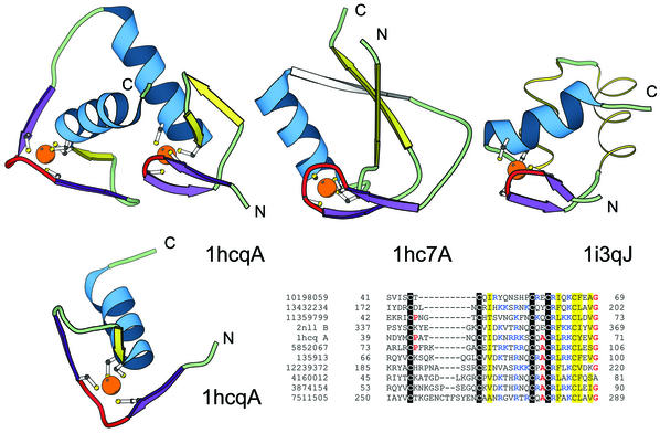
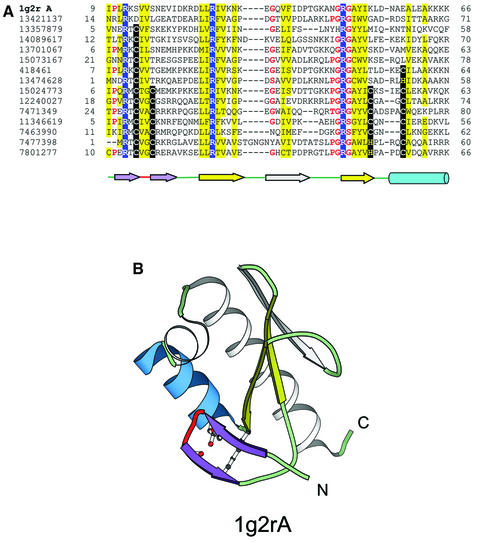
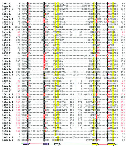
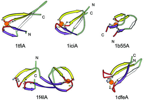
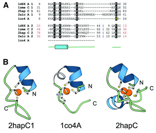
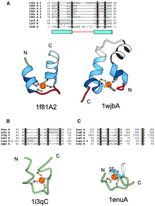
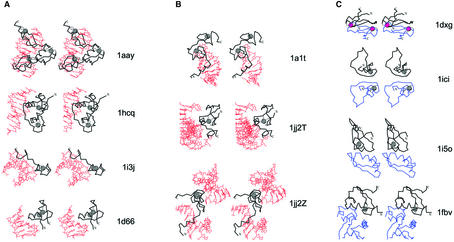
Zinc fingers are small protein domains in which zinc plays a structural role contributing to the stability of the domain. Zinc fingers are structurally diverse and are present among proteins that perform a broad range of functions in various cellular processes, such as replication and repair, transcription and translation, metabolism and signaling, cell proliferation and apoptosis. Zinc fingers typically function as interaction modules and bind to a wide variety of compounds, such as nucleic acids, proteins and small molecules. Here we present a comprehensive classification of zinc finger spatial structures. We find that each available zinc finger structure can be placed into one of eight fold groups that we define based on the structural properties in the vicinity of the zinc-binding site. Three of these fold groups comprise the majority of zinc fingers, namely, C2H2-like finger, treble clef finger and the zinc ribbon. Evolutionary relatedness of proteins within fold groups is not implied, but each group is divided into families of potential homologs. We compare our classification to existing groupings of zinc fingers and find that we define more encompassing fold groups, which bring together proteins whose similarities have previously remained unappreciated. We analyze functional properties of different zinc fingers and overlay them onto our classification. The classification helps in understanding the relationship between the structure, function and evolutionary history of these domains. The results are available as an online database of zinc finger structures.


Figures

Figure 1
Figure 1
Structure-based sequence alignment of the C2H2 finger fold group. For each sequence, the PDB entry name and chain ID (blank ID is replaced by A), fragment number (if more than one fragment is shown in the alignment), starting and ending residue numbers corresponding to the PDB numbering are given. Sequences in lower case italics are disordered in the structure. Regions in upper case italics differ in the secondary structure from the consensus secondary structure. Zinc ligands are boxed in black and non-zinc-binding residues at the same position are boxed in red. Zinc ligands in other sites are boxed in dark gray. Uncharged residues (all amino acids except D,E,K,R) in mostly hydrophobic sites are highlighted in yellow, non-hydrophobic residues (all amino acids except W,F,Y,M,L,I,V) at mostly hydrophilic sites are highlighted in light gray, small residues (G,P,A,S,C,T,V) at positions occupied by mostly small residues are shown in red letters. Charged residues K and R are marked in blue if they are present in the vicinity of a zinc-binding ligand. Long insertions are not displayed for clarity, and the number of omitted residues is specified in brackets. Regions of circular permutation are separated by a ‘|’ mark and the sequence number of the residues around the permuted region are shown in red. Sequences are further grouped into families. The same coloring and labeling scheme is used for figures of all fold groups of zinc fingers. Coloring unique to a particular figure is mentioned in its legend. A secondary structure consensus is shown below the alignment. β-strands are displayed as arrows and α-helices as cylinders. Color shading and labels of secondary structure elements correspond to those in the respective structural diagrams. Families are separated from each other by a larger spacing between the sequences. (A) Structure-based sequence alignment of domains of the C2H2 finger fold group. Here the domains are grouped into two families: the C2H2 like fingers (1ncsA, 1tf6A, 1zfdA, 1ubdC, 2gliA, 1tf6D, 1bhiA, 1sp2A, 1rmdA, 1aayA, 1znfA, 2adrA, 1sp1A, 1bboA, 2drpA, 1yuiA, 1ej6C, 1klrA, 1k2fA, 1fv5A and 1fu9A) and the IAP domains (1g73C, 1jd5A, 1c9qA and 1e31A). (B) Structure diagrams of the classical C2H2 zinc-binding motif (1tf6; chain D, residues 133–162) and BIR domain of Xiap-Bir3 domain of the human IAP (1g73 chain C, residues 296–330). The C2H2 motif found among many transcription factors is comprised of a zinc knuckle and a short β-hairpin at the N-terminus followed by a small loop and an α-helix. Two ligands each for zinc binding are contributed from the knuckle and the C-terminal part of the α-helix. The side chain of residues from the N-terminal part of the α-helix are involved in binding to the major groove of DNA in members of this class. The BIR domain, comprised of about 70 residues, has a conserved CCHC zinc-binding motif. The BIR domain is made up of a three-stranded β-sheet and four short α-helices. The colored region (non-gray) resembles a classic C2H2 motif, with two ligands each for zinc binding being contributed from a knuckle and the C-terminal part of an α-helix. The ribbon diagrams for this and all other diagrams in this manuscript were rendered by BOBSCRIPT (78,79), a modified version of MOLSCRIPT (24). (C) Alignment of representative protein sequences of IAP domains. The alignment includes apoptosis inhibitor survivin (1e31) and proteins from double-stranded DNA viruses Baculovirus (GI number 12597599, 1100734, 9631046, 1567532) and Iridovirus (GI number 1353180). The U-shaped transcription factor (1fu9) sequence is also included to better illustrate the resemblance between the IAP domains and the classical C2H2 fingers.
Figure 2
Figure 2
‘Gag knuckle’ fold group of zinc fingers. (A) Structural alignment of members of the ‘Gag knuckle’ fold group. We define three families for this fold group. Most of the members in the alignment are from the nucleocapsid protein of retroviruses (retroviral Gag knuckle family: 1a1tA, 1dsqA, 1dsvA, 1a6bB). A zinc-binding domain from the large subunit of RNA polymerase II (polymerase Gag knuckle family: 1i3qA) and from the reovirus outer capsid protein σ3 (reovirus outer capsid protein σ3 Gag knuckle family: 1fn9A) have been included in the alignment primarily based on structural similarities to the NC proteins of retroviruses. (B) Representative Gag knuckle from HIV nucleocapsid (1a1t, chain A, residue 33–52) and RNA polymerase II large subunit (1i3q, chain A, residue 64–83). The Gag knuckle is typically characterized by a zinc knuckle, which contributes two ligands and two more ligands contributed from both ends of a small helix or a loop.
Figure 3
Figure 3
Structure-based sequence alignment of the treble clef fold group of zinc fingers. Here the structures are grouped into 10 families. The families are: RING finger-like (1chcA, 1borA, 1jm7A, 1jm7B, 1rmdA, 1fbvA, 1g25A, 1ldjB, 1e4uA, 1dcqA); protein kinase cysteine-rich domain (1ptqA, 1faqA, 1kbeA, 1e53A); phosphatidylinositol-3-phosphate-binding domain (1vfyA, 1dvpA, 1jocA, 1zbdB, 1fp0A, 1f62A); nuclear receptor-like finger (1fjfN, 1jj2T, 1ee8A, 1k3wA, 1l2bA, 1ffyA, 1zfoA, 1i3jA, 1xpaA, 4gatA, 2gatA, 1gnfA, 1b8tA, 1imlA, 1g47A, 1hcqA, 1kb6A, 1lnrY); YlxR-like hypothetical cytosolic protein (1g2rA); prolyl tRNA synthetase (1hc7A); NAD+-dependent DNA ligase treble clef domain (1dgsA); YacG-like hypothetical protein (1lv3A); His-Me endonucleases (1en7A, 1bxiB, 1ql0A, 1a73A, 1mhdA); RPB10 protein from RNA polymerase II (1i3qJ, 1ef4A). The coloring scheme follows that in Figure 1.
Figure 4
Figure 4
Structural comparisons of representative treble clef fingers. Structural diagrams of the treble clef of the steroid hormone ‘estrogen’ receptor DNA-binding domain showing both zinc-binding sites (1hcq, chain A, residues 3–36 and 39–71), prolyl-tRNA synthetase (1hc7, chain A, residues 454–477; 418–440) and the RPB10 protein from RNA polymerase II (1i3q, chain J, residues 3–54) are shown to illustrate the wide range of variations in the structure of the treble clef finger. The N-terminal site of 1hcq is a typical treble clef motif. The C-terminal site (below), reoriented for clarity, lacks the β-hairpin shown in yellow for the N-terminal treble clef and has a distorted knuckle (red). This zinc-binding region shows some resemblance to other treble clef fingers in the placement of the ligands for zinc binding. However, the knuckle, which contributes the other two ligands in treble clef fingers, is significantly different and does not align structurally with those in other fingers. PSI-BLAST (33,34) alignment of representative nuclear receptor sequences. The sequences included in the alignment are: the nuclear factor NHR-63 (gi 10198059), peroxisome proliferator activated receptor (gi 13432234), NHR-18 (gi 11359799), retenoid X receptor (2nll), estrogen receptor (1hcq), tailless protein (TLL_DROME gi 135913), FTZ-F1 (gi 12239372), dissatisfaction (gi 4160012), NHR-67 (gi 3874154) and NHR-2 (gi 7511505). The gi accession number, start and end sequence number are given. The coloring scheme follows Figure 1.
Figure 5
Figure 5
Treble clef finger from the YlxR-like hypothetical cytosolic protein. (A) PSI-BLAST (33,34) alignment of representative homologs of the YlxR-like hypothetical cytosolic protein from the Nusa/Infb operon in S.pneumoniae. All protein sequences shown in this alignment are hypothetical proteins from bacterial sources and do not have any function assigned to them. Three conserved arginine residues are highlighted in blue. The secondary structure of 1g2r is shown below the alignment. β-Strands are displayed as arrows and α-helices are displayed as cylinders. The colors correspond to secondary structural elements shown in (B). The start and end residues of each sequence are given and the leftmost column contains the gi number of the sequence. (B) Structural diagram of the YlxR-like hypothetical cytosolic protein. This protein is a treble clef finger with an additional strand inserted between the second β-hairpin (yellow). Also the zinc-binding site of this protein is disintegrated and the structurally equivalent residues at the metal binding region are shown as ball-and-stick.
Figure 6
Figure 6
Structure-based sequence alignment of the zinc ribbon fold group. The proteins are grouped into nine families: classical zinc ribbon (1tfiA, 1qypA, 1i50I, 1pftA, 1dl6A, 1d0qA, 1yuaA, 1i50B, 1i50A, 1kjzA, 1i50L, 1l1oF, 1i5oD, 1qf8A, 1jj2Z, 1lnrZ, 1jj22, 1jj2Y, 1lnr1), the cluster binding domain of Rieske iron sulfur protein (1ezvE, 1rfsA, 1g8kB, 1eg9A, 1fqtA), the AdDBP zinc ribbons (1aduA), the B-box zinc finger (1freA), rubredoxin family (1dx8A, 1b71A, 1irnA, 1dxgA, 2occF), rubredoxin-like domains in enzymes (1f4lA, 1a8hA, 1ileA, 1gaxA, 1zinA, 1e4vA, 1zakA, 1iciA, 1ma3A, 1j8fA, 1gh9A), Btk motif (1b55A), ribosomal protein L36 (1dfeA) and the cysteine-rich domain of the chaperone protein DnaJ (1exkA). The coloring scheme follows Figure 1.
Figure 7
Figure 7
Structural diagrams of the zinc ribbons. Zinc ribbons from the transcriptional factor SII, C-terminal domain (1tfi, chain A, residues 8–50), Sir2 homolog—transcriptional regulatory protein (1ici, chain A, residues 141–156; 119–134), zinc-binding domain from Bruton’s tyrosine kinase (1b55, chain A, residues 161–166; 142–160), methionyl-tRNA synthetase (1f4l, chain A, site 1: residues 171–189; 122–139, site 2: residues 141–168) are shown. Different circular permutations are seen in the ribbon. The structure diagram of the zinc ribbon from the ribosomal protein L36 from T.thermophilus (1dfe, chain A, residues 7–37) is shown to illustrate the very different orientation of the two knuckles in the structure as compared to other zinc ribbons. The coloring follows Figure 1.
Figure 8
Figure 8
Zn2/Cys6 fold group of zinc fingers. (A) Structure-based sequence alignment of members of the Zn2/Cys6 zinc finger fold group. Members of this fold group contain two zinc-binding sites with the zinc ions coordinated by six cysteine ligands. The fourth ligand to the second site is the N-terminal cysteine. The members of the first family include the transcriptional regulatory proteins Gal4 (1d66A), Hap1 (2hapC), PUT3 (1zmeC), ethanol regulon transcriptional activator (2alcA). The second family contains a zinc domain conserved in yeast copper responsive transcription factors (1co4A). (B) Structural diagrams of the N-terminal zinc-binding site of the Zn2/Cys6 fingers from the Hap1 transcription factor (2hapC1, chain C, residues 61–83) and the zinc domain conserved in yeast copper responsive transcription factors (1co4A, chain A, residues 8–26). 1co4 has only one zinc-binding site unlike the members of Zn2/Cys6 finger family (2hapC, chain C, residues 61–99) that contain two sites. The second zinc-binding site shares two ligands with the first site. Coloring scheme follows that in Figure 1.
Figure 9
Figure 9
TAZ2 domain-like zinc finger fold group and short zinc-binding loops. (A) Structure-based sequence alignment of members of the TAZ2 domain-like zinc finger fold group. This fold group includes three families, TAZ2 domain family that consists of the structures of the three zinc-binding sites of CBP (1f81A, 1l8cA), the N-terminal zinc-binding domain of HIV-1 integrase (1wjbA) and the zinc-binding domain from DNA polymerase III γ subunit (1jr3A, 1jr3E). Structural diagrams of the second zinc-binding site of the CBP (1f81A2, chain A, residues 38–60) and the N-terminal zinc-binding domain of HIV-1 integrase (1wjbA, chain A, residues 36–48 and 12–20) are shown. Coloring scheme follows that in Figure 1. (B) Structural alignment of short zinc-binding loops present in large protein chains. The proteins in the alignment include loops from the human α alcohol dehydrogenase (1hsoA), sorbitol dehydrogenase (1e3jA), the 45 kDa polypeptide Rpb3 of the DNA-directed RNA polymerase II (1i3qC), the δ′ subunit of the clamp-loader complex of DNA polymerase III (1a5tA), intron encoded homing endonuclease I-PpoI (1cyqA) and core Gp32 ssDNA-binding protein (1gpcA). A representative figure of the RNA polymerase II protein Rpb3 (1i3qC, chain C, residue 84–97) is shown. (C) In some proteins the fourth ligand comes from a secondary structure far away in sequence from the other three ligands. This subgroup includes members from the tRNA-guanine transglycosylase (1enuA, 1iq8A), protein kinase domain of a Trp Ca-channel (1ia9A), intron encoded homing endonuclease I-PpoI (1cyqA), the Vsr endonuclease (1cw0A) and the RING finger protein Rbx1 (1ldjB). A representative figure of the zinc-binding region from tRNA-guanine transglycosylase (1enu, chain A, residue 316–325; 348–350) is shown. Three of the ligands of the zinc-binding site are from a loop and the fourth is from an α-helix.
Figure 10
Figure 10
Functional properties of zinc fingers. Cα traces of the zinc finger containing proteins are displayed in black with the N- and the C-terminus labeled. Zinc ions are shown as gray balls and Fe2+ ions are shown in purple. Side chains of zinc ligands or residues in corresponding sites are shown in black. Cα traces of the polypeptide chains interacting with the zinc fingers are dark blue. Nucleic acid is colored in red. (A) Protein–DNA interactions. Stereo diagrams of the Zif268 zinc finger from the C2H2-like group (1aay), the estrogen receptor (1hcq) and the intron endonuclease I-Tevi (1i3j) that belongs to the treble clef fingers and the GAL4 protein (1d66), which is a Zn2/Cys6 finger, are shown to illustrate the modes of DNA binding by zinc fingers. (B) Protein–RNA interactions. Stereo diagrams of the structure of the Gag knuckle from the HIV-1 nucleocapsid protein (1a1t) and the ribosomal proteins L24E (chain T of 1jj2; treble clef) and L37E (chain Z of 1jj2; zinc ribbon) interacting with RNA. (C) Protein–protein interactions. Stereo diagrams of desulforedoxin dimer (1dxg) and the homodimer of the zinc-binding domain of the Sir-2 homology protein (1ici). Also shown are the heterodimer of the zinc ribbon from E.coli aspartate transcarbamoylase regulatory chain (1i5o chain D; black) with the catalytic chain (1i5o chain C; blue) and the treble clef RING finger domain of the signal transduction protein Cbl in complex with ubiquitin-conjugating enzyme Ubch7 (1fbv chains A and C).

References

    1. Berman H.M., Battistuz,T., Bhat,T.N., Bluhm,W.F., Bourne,P.E., Burkhardt,K., Feng,Z., Gilliland,G.L., Iype,L., Jain,S. et al. (2002) The protein data bank. Acta Crystallogr. D Biol. Crystallogr., 58, 899–907. - PubMed
    1. Murzin A.G. (1998) How far divergent evolution goes in proteins. Curr. Opin. Struct. Biol., 8, 380–387. - PubMed
    1. Grishin N.V. (2001) Treble clef finger—a functionally diverse zinc-binding structural motif. Nucleic Acids Res., 29, 1703–1714. - PMC - PubMed
    1. Holm L. and Sander,C. (1997) Dali/FSSP classification of three-dimensional protein folds. Nucleic Acids Res., 25, 231–234. - PMC - PubMed
    1. Shindyalov I.N. and Bourne,P.E. (1998) Protein structure alignment by incremental combinatorial extension (CE) of the optimal path. Protein Eng., 11, 739–747. - PubMed