. 2003 Jan 15;31(2):562-9.
doi: 10.1093/nar/gkg152.

Basal transcriptional regulation of human damage-specific DNA-binding protein genes DDB1 and DDB2 by Sp1, E2F, N-myc and NF1 elements

Affiliations

Basal transcriptional regulation of human damage-specific DNA-binding protein genes DDB1 and DDB2 by Sp1, E2F, N-myc and NF1 elements

Anne F Nichols et al. Nucleic Acids Res. .

Abstract

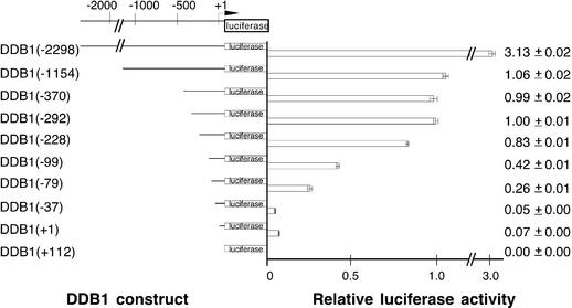
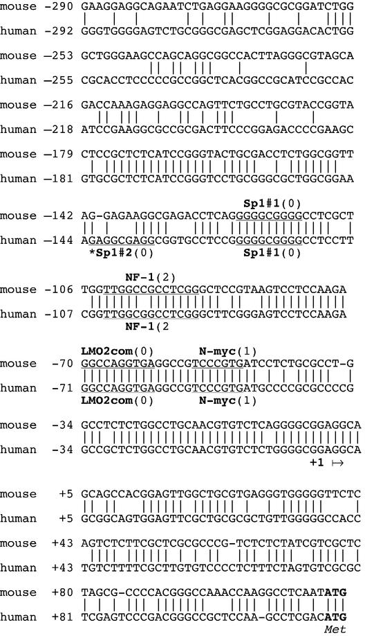
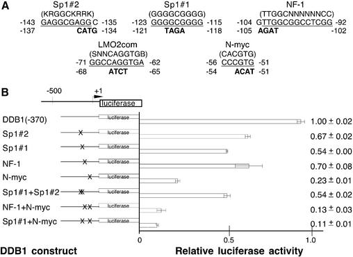
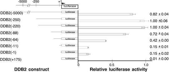
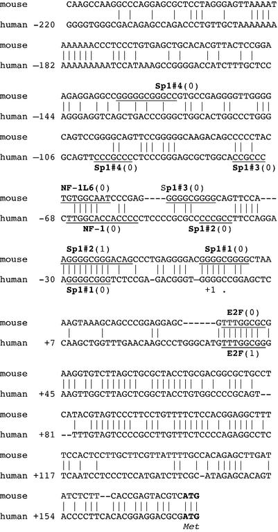
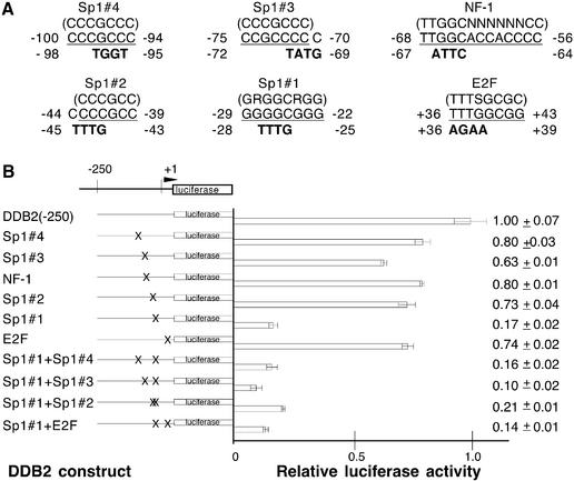
The human DDB1 and DDB2 genes encode the 127 and 48 kDa subunits, respectively, of the damage-specific DNA-binding protein (DDB). Mutations in the DDB2 gene have been correlated with the hereditary disease xeroderma pigmentosum group E. We have investigated the proximal promoters of the DDB genes, both of which are G/C-rich and do not contain a TATA box. Transient expression analysis in HeLa cells using a luciferase reporter system indicated the presence of core promoters located within 292 bp (DDB1) and 220 bp (DDB2) upstream of the putative transcription initiation sites. Both core promoters contain multiple active Sp1 sites, with those of DDB1 at -123 to -115 and of DDB2 at -29 to -22 being critical determinants of promoter activity. In addition, an N-myc site at -56 to -51 for DDB1 is an essential transcription element, and mutations in a DDB1 NF-1 site at -104 to -92, a DDB2 NF-1 site at -68 to -56 and a DDB2 E2F site at +36 to +43 also reduce promoter activity. Taken together, these results suggest a regulation of basal transcription typical of cell cycle-regulated genes, and therefore support conjectures that the DDB heterodimer and/or its subunits have functions other than direct involvement in DNA repair.


Figures

Figure 1
Figure 1
Transcription activities in log phase HeLa cells after deletions within the upstream region of the DDB1 gene. A series of 5′-unidirectional deletions were fused in-frame with the luciferase reporter gene and analyzed by transient transfection as described in Materials and Methods. Luciferase activities were normalized to β-galactosidase activity, which was also measured to control for transfection efficiency. Less than a 2-fold variation in transfection efficiency was observed. At least four extracts from two or more independent transfections were evaluated for each construct. The construct number in parentheses indicates the length of the tested promoter region upstream of the putative transcription initiation site (designated by the bent arrow at +1). Luciferase activity is shown relative to DDB1(–292). Means and standard deviations are indicated to the right.
Figure 2
Figure 2
Nucleotide sequence and potential regulatory elements of the promoter region of the DDB1 gene in mouse and human genomic DNAs. The consensus sequences of the potential transcription factor-binding sites are underlined. Numbers in parentheses are the number of mismatches as evaluated with the TRANSFAC database consensus sequence. Elements which were tested, although not found in both mouse and human DNAs, are marked by an asterisk. The putative transcription initiation site (+1) is indicated by an arrow, and ATG is the translation initiation site.
Figure 3
Figure 3
Mutational analysis of putative proximal promoter transcription elements of the DDB1 gene. (A) The wild-type sequences that were analyzed by mutation are shown with their nucleotide position in the DDB1 gene. The consensus sequences are shown above in parentheses, the DDB1 wild-type element is underlined and mutations that were introduced are indicated below the wild-type sequence in bold. Mutations were introduced by overlapping PCR into the DDB1(–370) plasmid. (B) Potential transcription elements that were mutated are indicated to the left and correspond to those in (A). X on the construct schematic indicates the location of the mutation. The plasmids were transfected into log phase HeLa cells and luciferase activity was measured in cell extracts and standardized to β-galactosidase activity, which was also measured to control for transfection efficiency. Less than a 2-fold variation in transfection efficiency was observed. At least four extracts from two or more independent transfections were evaluated for each construct. Luciferase activity is shown relative to the control wild-type DDB1(–370). Means and standard deviations are indicated to the right.
Figure 4
Figure 4
Transcription activities in log phase HeLa cells after deletions within the upstream region of the DDB2 gene. A series of 5′-unidirectional deletions were fused in-frame with the luciferase reporter gene and analyzed by transient transfection as described in Materials and Methods. Luciferase activity was standardized to β-galactosidase activity to control for variations in transfection activity. Less than a 2-fold variation in transfection efficiency was observed. At least four extracts from two or more independent transfections were evaluated for each construct. The construct number in parentheses indicates the length of the tested promoter region upstream of the putative transcription initiation site (designated by the bent arrow at +1). Luciferase activity is shown relative to DDB1(–220). Means and standard deviations are indicated to the right.
Figure 5
Figure 5
Nucleotide sequence and potential regulatory elements of the promoter region of the DDB2 gene in mouse and human genomic DNAs. The consensus sequences of the potential transcription factor-binding sites are underlined. Numbers in parentheses are the number of mismatches as evaluated with the TRANSFAC database consensus sequence. Elements which were tested, although not found in both mouse and human DNAs, are marked by an asterisk. The putative transcription initiation site (+1) is indicated by an arrow, and ATG is the translation initiation site.
Figure 6
Figure 6
Mutational analysis of putative proximal promoter transcription elements of the DDB2 gene. (A) The wild-type sequences that were analyzed by mutation are shown with their nucleotide position in the DDB2 gene. The consensus sequences are shown above in parentheses, the DDB2 wild-type element is underlined and mutations that were introduced are indicated below the wild-type sequence in bold. Mutations were introduced by overlapping PCR into the DDB2(–250) plasmid. (B) Potential transcription elements that were mutated are indicated to the left and correspond to those in (A). X on the construct schematic indicates the location of the mutation. The plasmids were transfected into log phase HeLa cells and luciferase activity was measured in cell extracts and standardized to β-galactosidase activity, which was also measured to control for transfection efficiency. Less than a 2-fold variation in transfection efficiency was observed. At least four extracts from two or more independent transfections were evaluated for each construct. Luciferase activity is shown relative to the control wild-type DDB2(–250). Means and standard deviations are indicated to the right.

References

    1. Cleaver J.E. and Kraemer,K.H. (1995) Xeroderma pigmentosum and Cockayne Syndrome. In Scriver,C.R., Beaudet,A.L., Sly,W.S. and Valle,D. (eds), The Metabolic and Molecular Bases of Inherited Disease, Vol. III. McGraw-Hill, New York, NY, pp. 4393–4419.
    1. Chu G. and Chag,E. (1988) Xeroderma pigmentosum group E cells lack a nuclear factor that binds to damaged DNA. Science, 242, 564–567. - PubMed
    1. Kataoka H. and Fujiwara,Y. (1991) UV-damage-specific DNA-binding protein in xeroderma pigmentosum complementation group E. Biochem. Biophys. Res. Commun., 175, 1139–1143. - PubMed
    1. Keeney S., Wein,H. and Linn,S. (1992) Biochemical heterogeneity in xeroderma pigmentosum complementation group E. Mutat. Res., 273, 49–56. - PubMed
    1. Otrin V.R., Kuraoka,I., Nardo,T., McLenigan,M., Eker,A.P.M., Stefanini,M., Levine,A.S. and Wood,R.D. (1998) Relationship of the xeroderma pigmentosum group E defect to chromatin and DNA binding proteins UV-DDB and replication protein A. Mol. Cell. Biol., 18, 3182–3190. - PMC - PubMed

Publication types

MeSH terms