. 2003 Jan;13(1):55-63.
doi: 10.1101/gr.791403.

Pericentromeric duplications in the laboratory mouse

Affiliations

Pericentromeric duplications in the laboratory mouse

James W Thomas et al. Genome Res. 2003 Jan.

Abstract

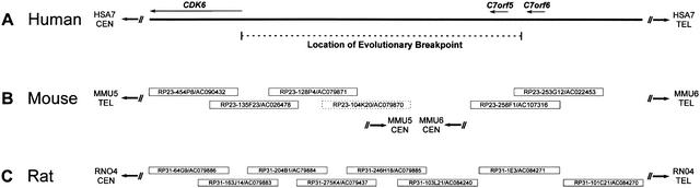
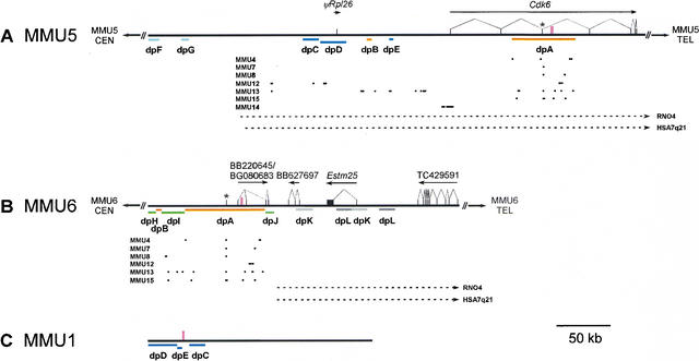
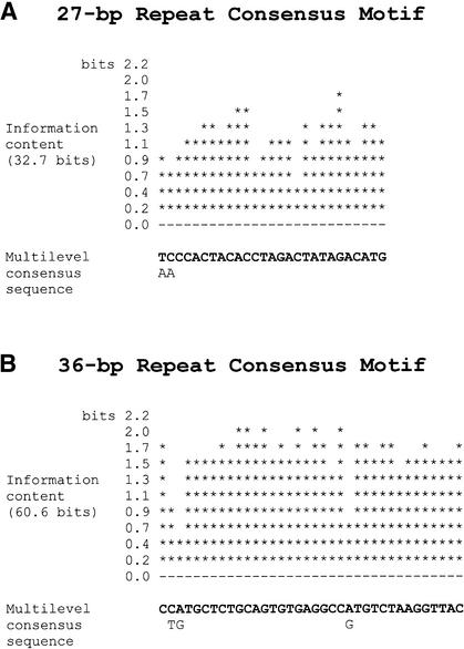
Duplications have long been postulated to be an important mechanism by which genomes evolve. Interspecies genomic comparisons are one method by which the origin and molecular mechanism of duplications can be inferred. By comparative mapping in human, mouse, and rat, we previously found evidence for a recent chromosome-fission event that occurred in the mouse lineage. Cytogenetic mapping revealed that the genomic segments flanking the fission site appeared to be duplicated, with copies residing near the centromere of multiple mouse chromosomes. Here we report the mapping and sequencing of the regions of mouse chromosomes 5 and 6 involved in this chromosome-fission event as well as the results of comparative sequence analysis with the orthologous human and rat genomic regions. Our data indicate that the duplications associated with mouse chromosomes 5 and 6 are recent and that the resulting duplicated segments share significant sequence similarity with a series of regions near the centromeres of the mouse chromosomes previously identified by cytogenetic mapping. We also identified pericentromeric duplicated segments shared between mouse chromosomes 5 and 1. Finally, novel mouse satellite sequences as well as putative chimeric transcripts were found to be associated with the duplicated segments. Together, these findings demonstrate that pericentromeric duplications are not restricted to primates and may be a common mechanism for genome evolution in mammals.


Figures

Figure 1.
Figure 1.
Physical mapping of the human, mouse, and rat genomic regions flanking an evolutionary breakpoint. The depicted segment of HSA7q21 (A) and the orthologous region of RNO4 (C) are contiguous in each species; however, the orthologous segment in the mouse genome is split between two regions (B), with portions located at the proximal (i.e., centromeric) ends of MMU5 and MMU6. The indicated location of the evolutionary breakpoint on HSA7q21 is based on genetic and cytogenetic mapping studies (Thomas et al. 1999). The positions of the three genes (CDK6, C7orf5, and C7orf6) on HSA7q21 are based on finished genomic sequence (GenBank nos. , , , , and ). Also shown are the minimal tiling paths of BACs selected for sequencing the orthologous regions on MMU5, MMU6, and RNO4 (Thomas et al. 1999; Summers et al. 2001), with the clone name/GenBank accession number indicated for each. Note that the mouse BACs depicted here are the most proximal clones in larger (20-BAC) tiling paths assembled for both MMU5 and MMU6 (data not shown). One clone (RP23–104K20) was subsequently found not to map to MMU5 (indicated by a dashed box). The orientation of the depicted sequences and clones relative to each telomere (TEL) and centromere (CEN) is indicated. Note that the human sequence as well as the mouse and rat clones are not drawn to scale.
Figure 2.
Figure 2.
Annotated features of the MMU5, MMU6, and MMU1 sequences. The generated sequences from MMU5 (A), MMU6 (B), and MMU1 (C) were compiled and annotated as described (see Methods), with the corresponding annotated sequence files available at www.nisc.nih.gov/data. The positions and intron-exon structures of the indicated genes were determined (arrows indicate the direction of transcription and black rectangles represent exons). Also indicated are the positions of satellite sequences (tall pink boxes) and the location of Cdk6 exon 3 (designated by *). The various colored lines labeled dpA through dpL depict the relative positions of duplicated segments (see Table 1). The positions of aligned, near-identical BAC-end sequences are represented by dots, with each sorted based on the mouse chromosome from which the originating BAC was mapped. The portions of the MMU5 and MMU6 sequences orthologous to RNO4 and HSA7q21 are denoted by the dashed arrows. The orientation of the MMU5 and MMU6 sequences relative to each telomere (TEL) and centromere (CEN) is indicated; note that the orientation of the MMU1 sequence relative to the centromere and telomere could not be determined. Additional details about the various annotated features are provided in the text.
Figure 3.
Figure 3.
Consensus sequences of novel mouse satellites. (A) Consensus sequence of the 27-bp satellite derived by MEME analysis of sequence flanking dpE on MMU1. The consensus represents 83 monomers, each 27 bp in length and occurring in a tandem, head-to-tail fashion within BAC RP23–104K20. The information content (in bits) is included to indicate the degree of conservation at each position in the consensus. The base with the greatest probability of occurrence at each position is shown in the first line of the multilevel consensus sequence; alternative bases are indicated on the second line only if they occur with a probability greater than 0.2. (B) Consensus sequence of the 36-bp satellite derived by MEME analysis of sequence within dpA. The consensus represents 106 monomers, each 36 bp in length, derived from several genomic locations: 62 monomers from MMU5, 40 from MMU6, and four from MMU8. Monomers of this 36-bp satellite are arranged in a tandem, head-to-tail orientation.

References

    1. Bailey J.A., Yavor, A.M., Massa, H.F., Trask, B.J., and Eichler, E.E. 2001. Segmental duplications: Organization and impact within the current Human Genome Project assembly. Genome Res. 11: 1005-1017. - PMC - PubMed
    1. Bailey J.A., Yavor, A.M., Viggiano, L., Misceo, D., Horvath, J.E., Archidiacono, N., Schwartz, S., Rocchi, M., and Eichler, E.E. 2002. Human-specific duplication and mosaic transcripts: The recent paralogous structure of chromosome 22. Am. J. Hum. Genet. 70: 83-100. - PMC - PubMed
    1. Bailey T. and Elkan, C., 1994. Fitting a mixture model by expectation maximization to discover motifs in biopolymers. In Conference on intelligent systems for molecular biology, pp. 28–36. AAAI Press, Menlo Park, CA. - PubMed
    1. Dehal P., Predki, P., Olsen, A.S., Kobayashi, A., Folta, P., Lucas, S., Land, M., Terry, A., Ecale Zhou, C.L., Rash, S., et al. 2001. Human chromosome 19 and related regions in mouse: Conservative and lineage-specific evolution. Science 293: 104-111. - PubMed
    1. Eichler E.E. 2001. Recent duplication, domain accretion and the dynamic mutation of the human genome. Trends Genet. 17: 661-669. - PubMed

Publication types

LinkOut - more resources