. 2003 Feb;23(3):815-25.
doi: 10.1128/MCB.23.3.815-825.2003.

Recruitment of a repressosome complex at the growth hormone receptor promoter and its potential role in diabetic nephropathy

Affiliations

Recruitment of a repressosome complex at the growth hormone receptor promoter and its potential role in diabetic nephropathy

P M Gowri et al. Mol Cell Biol. 2003 Feb.

Abstract

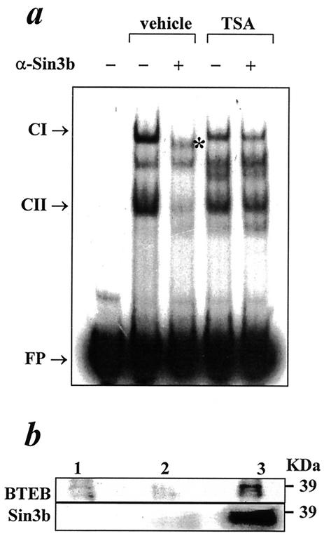
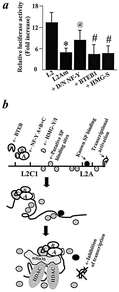
The growth hormone (GH)-GH receptor (GHR) axis modulates growth and metabolism and contributes to complications of diabetes mellitus. We analyzed the promoter region of the dominant transcript (L2) of the murine GHR to determine that a cis element, L2C1, interacts with transcription factors NF-Y, BTEB1, and HMG-Y/I. These proteins individually repress GHR expression and together form a repressosome complex in conjunction with mSin3b. The histone deacetylase inhibitor trichostatin A increases expression of the murine GHR gene, enhances association of acetyl-H3 at L2C1, inhibits formation of the repressosome complex, and decreases NF-Y's association with L2C1. Our studies reveal that murine models of experimental diabetes mellitus are characterized by reduced hepatic GHR expression, decreased acetyl-H3 associated with L2C1, and increased formation of the repressosome complex. In contrast, in the kidney diabetes mellitus is associated with enhanced GHR expression and lack of alteration in the assembly of the repressosome complex, thus permitting exposure of kidneys to the effects of elevated levels of GH in diabetes mellitus. Our findings define a higher-order repressosome complex whose formation correlates with the acetylation status of chromatin histone proteins. The delineation of the role of this repressosome complex in regulating tissue-specific expression of GHR in diabetes mellitus provides a molecular model for the role of GH in the genesis of certain microvascular complications of diabetes mellitus.


Figures

FIG. 1.
FIG. 1.
Identification of the cis element, L2C1. (a) Transient expression analyses of the promoter-regulatory region of L2 transcript of the murine GHR. Luciferase expression plasmids containing various portions of the 5′-flanking sequence of L2 transcript were transiently transfected in BNL CL.2 cells and assayed for luciferase activity. The arrow indicates the orientation of GHR DNA relative to the direction of GHR gene transcription. Results represent means ± standard errors. Using analysis of variance, P < 0.05 in comparison with pGL3B (*) and pGL3B-L2[-344] (#). (b) DNA footprint analysis. 32P-single-end-labeled DNA fragment (generated by PCR) was incubated with 20 μg of nuclear extract from adult mouse liver (lanes 2 and 3) or no protein (lane 1) and tested for the presence of DNase I footprint by the addition of DNase I (0.5 ng [lane 2] and 2 ng [lane 3]). Arrowheads indicate the protected bands within the footprinted region. The sequence of the 72-bp footprinted region is indicated with the putative GC (double-underlined) and CCAAT (single-underlined) boxes.
FIG. 2.
FIG. 2.
Identification of the proteins interacting at L2C1. (a) Nuclear proteins from mouse liver interact with the L2C1 site in the promoter of the L2 transcript of the murine GHR gene. 32P-labeled L2C1 was incubated with (lanes 1 to 3) or without (lane P) nuclear extracts prepared from liver tissues of adult mice in the absence (lane 1) or presence of antibodies against the NF-YA (lane 2) or BTEB1 (lane 3), electrophoresed, and subjected to autoradiography. The bands representing specific DNA-protein complexes (CI, CII), supershifted complexes (SS), and the free probe (FP) are indicated. (b) NF-YA and BTEB1 interact with L2C1. 32P-labeled L2C1 was incubated with nuclear extract from adult mouse liver without (lane NC) or with excess unlabeled oligonucleotide (lanes a [25×], b [50×], and c [100×]). The unlabeled oligonucleotide used was either the wild-type sequence (L2C1) or oligonucleotides with mutations in the NF-Y (NF-Ym), BTEB1 (BTEBm), or both (L2C1m) binding sites. The bands representing specific DNA-protein complexes (CI, CII) and the free probe (FP) are indicated. (c) HMG Y/I is a component of the CI complex. 32P-labeled L2C1 was incubated with (lanes 1 to 6) or without (lane P) nuclear extracts prepared from liver tissues from adult mice in the presence of either poly(dI-dC) · poly(dI-dC) (0 μg [lane 1], 1 μg [lane 2], and 2 μg [lane 3]) or anti-HMG-Y/I antibody (20 ng [lane 4], 40 ng [lane 5], 100 ng [lane 6]). The bands representing specific DNA-protein complexes (CI, CII) and the free probe (FP) are indicated. (d) L2C1 binds HMG Y/I. 32P-labeled L2C1 was incubated without (lane P) or with recombinant HMG-Y/I protein (lane 1), electrophoresed, and subjected to autoradiography. The bands representing L2C1-HMG complex (◂) and the unbound 32P-labeled L2C1 (FP) are indicated.
FIG. 3.
FIG. 3.
L2C1 represses the activity of the murine GHR gene promoter. (a) Transient transfection of luciferase reporter plasmids whose expression was controlled by wild-type GHR promoter (L2) or GHR promoter with a mutation designed to abolish the L2C1 binding sites for NF-Y, HMG-Y/I, and BTEB1 (L2C1 mutant) in BNL CL.2 cells. Results represent means ± SD of three or four independent transfections performed in triplicate. For luciferase activity, P < 0.05 compared to that of the empty vector (pGL3B) (*) or the wild-type GHR promoter (#), respectively. (b) NF-Y, BTEB1, and HMG-Y/I individually repress transcriptional activation. Transient cotransfection assays in BNL CL.2 cells with L2 luciferase reporter plasmid with constructs expressing the dominant negative form of NF-YA (DN/NF-Y, 250 ng), sense HMG-Y/I (HMG-S, 250 ng), or antisense HMG-Y/I (HMG-AS, 250 ng). Results represent means ± SD of three or four independent transfections performed in triplicate. *, P < 0.05 (luciferase activity compared to that of the wild-type GHR promoter); ns, statistically not significantly different from DN/NF-Y. (c) BNL CL.2 cells transiently cotransfected with the L2C1m-GHR reporter construct and expression plasmid for either D/N NF-Y, BTEB1, or sense HMG-Y/I. Results represent means ± SD of three or four independent transfections performed in triplicate. (d) Cotransfection of BTEB1 (500 ng) expression plasmid with luciferase constructs whose expression was controlled by wild-type promoter or GHR promoter with mutated NF-Y+HMG-Y/I or BTEB binding sites at L2C1. Results represent means ± SD of three or four independent transfections performed in triplicate. *, P < 0.05 in comparison to respective transfections carried out without BTEB1 (control).
FIG. 4.
FIG. 4.
Role of chromatin in modulating the activity of L2C1. (a) Effect of TSA exposure on BNL CL.2 cells stably transfected with L2- and -mCCAAT reporter constructs. Results represent mean ± SD of three independent experiments. *, P < 0.05 in comparison to the effect of TSA on cells stably transfected with an intact L2C1 site. (b) Fluorescent 5′-nuclease (TaqMan) real time RT-PCR analysis of expression of endogenous L2 transcript after exposure of BNL CL.2 cells to vehicle (alcohol) or TSA. The results are depicted as mean and range. *, P < 0.05, in comparison to the cells exposed to vehicle only. (c) 32P-labeled L2C1 was incubated with or without nuclear extracts prepared from BNL CL.2 cells exposed to vehicle or TSA in the absence or presence of antibodies against the NF-YA or BTEB1, electrophoresed, and subjected to autoradiography. The inset shows total NF-Y content in these cells as ascertained by EMSA with 32P-labeled NF-Y consensus oligonucleotide. The bands representing specific DNA-protein complexes (CI, CII), supershifted complexes (SS), and the free probe (FP) are indicated. (d) ChIP assay on BNL CL.2 cells treated without (vehicle) and with TSA. Immunoprecipitation with anti-NF-YA (top panel) and anti-acetyl-H3 (middle panel) antibodies. The input DNA and the sample without addition of the antibody (unbound) are also shown. The linearity of the PCR assay was established by assaying serial concentrations of the input DNA (bottom panel).
FIG. 5.
FIG. 5.
Protein-protein interaction at L2C1. (a) EMSA to demonstrate association of Sin3b with proteins interacting at L2C1. 32P-labeled L2C1 was incubated without and with nuclear extracts from vehicle or TSA-treated BNL CL.2 cells, in the absence or presence of antibody directed against Sin3b. The bands representing specific DNA-protein complexes (CI, CII), the band with altered electromobility (*), and the free probe (FP) are indicated. (b) Immunoblots to study in vivo interaction between NF-Y and mSin3b and NF-Y and BTEB. (Upper panel) Proteins from untransfected BNL CL.2 cells (lane 1) or BNL CL.2 cells transfected with BTEB1 expression plasmid (lanes 2 and 3) were immunoprecipitated with immunoglobulin G (IgG) (lane 2) or anti-BTEB antibody (lanes 1 and 3) and immunoblotted with anti-NF-YA antibody. The position of the molecular weight standard is indicated. (Lower panel) Total protein from BNL CL.2 cells not subjected to immunoprecipitation (lane 1) or immunoprecipitated with IgG (lane 2) or anti-Sin3b antibody (lane 3) and immunoblotted with anti-NF-YA antibody. The position of the molecular weight standard is indicated.
FIG. 6.
FIG. 6.
Mode of action of the repressosome. (a) Interaction of the repressosome complex with the minimal promoter. BNL CL.2 cells were transiently cotransfected with a wild-type GHR promoter construct containing an intact (L2) or mutated L2A (L2Am) site, and the latter was cotransfected with expression plasmids for D/N NF-Y (250 ng), HMG-Y/I (250 ng), or BTEB1(500 ng). Results represent means ± SD of three or four independent transfections performed in triplicate. *, P < 0.05 in comparison to luciferase activity of L2; P < 0.05 (@) or difference not significant (#) for luciferase activity compared to L2Am. (b) Model depicting the role of factors binding to the L2C1 site in the modulation of activity of the L2 promoter. Sp factors activate transcription of the GHR gene by interacting with GC boxes (such as the L2A site) in the minimal promoter of the L2 transcript. Binding of NF-Y/HMG-Y/I and BTEB1 at L2C1 results in DNA bending. Sin3b associates with BTEB and NF-Y and recruits histone deacetylases (HDACs) to the DNA, compacting the DNA. Changes in chromatin architecture brought upon by the repressosome result in loss of effect of the Sp factors at the L2A site and consequent inhibition of GHR transcription.
FIG. 7.
FIG. 7.
Increased binding activity of NF-Y to the L2C1 cis element in DM. (a) EMSA with liver nuclear extracts from nondiabetic (lanes 1 to 4) or diabetic (lanes 5 to 10) NOD mice. The probes used were L2C1 (upper panel), OCT-1 (middle panel), or NF-Y consensus binding site (lower panel). The densitometry of the blots is represented with the data normalized to the nondiabetic group. (b) ChIP assays were conducted with liver from nondiabetic and diabetic (STZ induced) mice and L2C1 detected by PCR. Immunoprecipitation with anti-acetyl-H3 (top panel) and anti-NF-YA (bottom panel). The input DNA and the sample without addition of the antibody (unbound) are also shown.
FIG. 8.
FIG. 8.
Role of L2C1 in regulation of GHR expression in kidney in IDDM. (a) Fluorescent 5′-nuclease (TaqMan) real-time RT-PCR analysis of expression of L2 transcript in liver and kidney in murine experimental DM. The results (mean and range) are depicted relative to nondiabetic (control) mice. *, P < 0.05 in comparison to control. (b) 32P-labeled L2C1 was incubated with (lanes 1 to 3) or without (lane P) nuclear extracts prepared from kidney of adult male mice in the absence (lane 1) or presence of antibodies against the NF-YA (lane 2) or BTEB1 (lane 3), electrophoresed, and subjected to autoradiography. (Inset) Overexposure of the autoradiograph to illustrate the supershift of C1 complex by NF-YA antibody. The bands representing specific DNA-protein complexes (CI, CII), supershifted complexes (SS), and the free probe (FP) are indicated. (c) Lack of alteration in binding of proteins at the L2C1 complex in IDDM. EMSA was carried out with kidney nuclear extracts from nondiabetic or diabetic mice. The probes used were L2C1 (upper panel) or NF-Y consensus binding site (lower panel). (d) ChIP assays with immunoprecipitation with anti-acetyl H3, anti-HMG, and anti-NF-YA were conducted on kidney tissues from nondiabetic and diabetic (STZ induced) mice and L2C1 detected by PCR. The input DNA and the sample without addition of antibody (unbound) are also shown.

References

    1. Baxter, R. C., J. M. Bryson, and J. R. Turtle. 1980. Somatogenic receptors of rat liver: regulation by insulin. Endocrinology 107:1176-1181. - PubMed
    1. Bellush, L. L., S. Doublier, A. N. Holland, L. J. Striker, G. E. Striker, and J. J. Kopchick. 2000. Protection against diabetes-induced nephropathy in growth hormone receptor/binding protein gene-disrupted mice. Endocrinology 141:163-168. - PubMed
    1. Caretti, G., M. C. Motta, and R. Mantovani. 1999. NF-Y associates with H3-H4 tetramers and octamers by multiple mechanisms. Mol. Cell. Biol. 19:8591-8603. - PMC - PubMed
    1. Cheung, P., C. D. Allis, and P. Sassone-Corsi. 2000. Signaling to chromatin through histone modifications. Cell 103:263-271. - PubMed
    1. Currie, R. A. 1997. Functional interaction between the DNA binding subunit trimerization domain of NF-Y and the high mobility group protein HMG-I(Y). J. Biol. Chem. 272:30880-30888. - PubMed

Publication types

MeSH terms