. 2003 Feb;23(3):1004-13.
doi: 10.1128/MCB.23.3.1004-1013.2003.

Transcriptional coactivation of bone-specific transcription factor Cbfa1 by TAZ

Affiliations

Transcriptional coactivation of bone-specific transcription factor Cbfa1 by TAZ

Cai Bin Cui et al. Mol Cell Biol. 2003 Feb.

Abstract

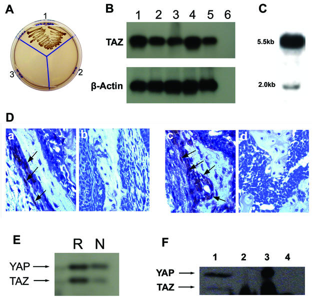
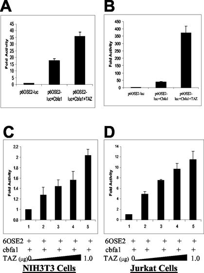
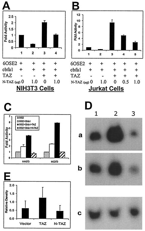
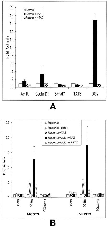
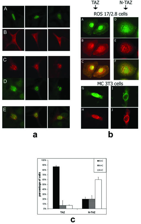
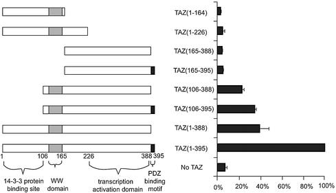
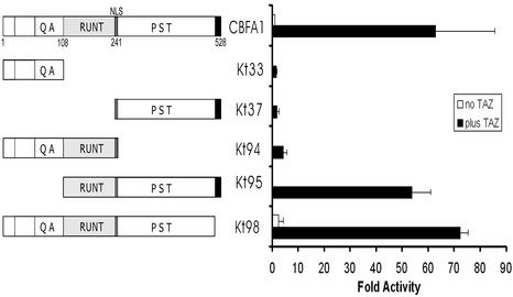
Core-binding factor 1 (Cbfa1; also called Runx2) is a transcription factor belonging to the Runt family of transcription factors that binds to an osteoblast-specific cis-acting element (OSE2) activating the expression of osteocalcin, an osteoblast-specific gene. Using the yeast two-hybrid system, we identified a transcriptional coactivator, TAZ (transcriptional coactivator with PDZ-binding motif), that binds to Cbfa1. A functional relationship between Cbfa1 and TAZ is demonstrated by the coimmunoprecipitation of TAZ by Cbfa1 and by the fact that TAZ induces a dose-dependent increase in the activity of osteocalcin promoter-luciferase constructs by Cbfa1. A dominant-negative construct of TAZ in which the coactivation domains have been deleted reduces osteocalcin gene expression down to basal levels. NIH 3T3, MC 3T3, and ROS 17/2.8 cells showed the expected nuclear localization of Cbfa1, whereas TAZ was distributed throughout the cytoplasm with some nuclear localization when transfected with either Cbfa1 or TAZ. Upon cotransfection by both Cbfa1 and TAZ, the transfected TAZ shows predominant nuclear localization. The dominant-negative construct of TAZ shows minimal nuclear localization upon cotransfection with Cbfa1. These data indicate that TAZ is a transcription coactivator for Cbfa1 and may be involved in the regulation of osteoblast differentiation.


Figures

FIG. 1.
FIG. 1.
(A) Two-hybrid analysis for the specificity of Cbfa1-TAZ interactions. Plasmid DNA from bait Cbfa1 was cotransformed with the prey plasmid containing TAZ cDNA into yeast strain AH 109. Section 1 is the cotransformed mixture, section 2 is the bait alone, and section 3 is the prey alone. Selection was made by growth on SD/−Ade/−His/−Leu/−Trp plates. Note the selective growth only in Section 1. (B) RT-PCR analysis of TAZ and β-actin in 7-day-old mouse pup tissues. Lanes: 1, calvaria; 2, long bones; 3, heart; 4, skin; 5, brain; 6, negative control PCR (i.e., PCR with no RT product). (C) Northern blot of total RNA from 7-day-old mouse calvaria hybridized with cDNA probe for mouse TAZ. Note the prominent 5.5-kb band, along with the less-prominent 2.0-kb band. (D) Immunohistochemistry of developing bone sections from 7-day-old mouse mandible (a and b) and femur (c and d). Paraffin-embedded sections were stained with rabbit antiserum to mouse TAZ (a and c) or normal rabbit serum (b and d). Note the staining of the osteoblast-like cells with the antiserum to TAZ (arrowheads). (E) Western blot of cell extract from ROS 17/2.8 (R) and NIH 3T3 cells (N) analyzed with antibody to mouse TAZ. Note the cross-reactivity to mouse YAP and the weaker expression of TAZ in NIH 3T3 cells compared to ROS 17/2.8 cells. (F) Western blot of immunoprecipitated proteins with antibody to Cbfa1 and Western blotting with antibody to mouse TAZ. Lanes: 1, total lysate from ROS 17/2.8 cells; 2, immunoprecipitation with antibody to Cbfa1; 3, immunoprecipitation with antibody to TAZ; 4, immunoprecipitation with nonspecific rabbit IgG. Note the immunoprecipitation of TAZ by Cbfa1 in lane 2. Interestingly, both TAZ and YAP are immunoprecipitated with antibody to mouse TAZ (lane 3).
FIG. 2.
FIG. 2.
(A and B) Transcriptional coactivation assay in NIH 3T3 (A) and Jurkat (B) cells. The results are expressed as fold increases over the control (p6OSE2-luc alone). A total of 1 μg of each construct was used for the NIH 3T3 cells, and 0.5 μg of each construct was used for the Jurkat cells. (C and D) Transcriptional coactivation assay in NIH 3T3 (C) and Jurkat (D) cells demonstrating the dose-responsive effects of TAZ. The results are expressed as the fold increases over p6OSE2-luc+Cbfa1 (positive control). p6OSE2-luc and Cbfa1 were used at 1 μg/well for NIH 3T3 cells and at 0.5 μg/well for Jurkat cells. The data in panels A and B are expressed as fold increases in activity compared to p6OSE2-luc alone, whereas the data in panels C and D are expressed as the fold increases in activity compared to p6OSE2-luc and Cbfa1. The differences in the fold activities expressed between panels A and B and panels C and D are noted.
FIG. 3.
FIG. 3.
(A and B) Effects of a dominant-negative construct of TAZ on osteoblast-specific gene expression. Transcriptional coactivation assays in NIH 3T3 (A) and Jurkat (B) cells show the dominant-negative effects of N-TAZ (truncated at the end of WW domain). The results are expressed as fold increases over p6OSE2-luc+Cbfa1 (positive control). p6OSE2-luc, Cbfa1, and TAZ were used at 1 μg/well for NIH 3T3 cells and at 0.5 μg/well for Jurkat cells. (C) Transcriptional coactivation assay in NIH 3T3 and MC 3T3 E1 cells performed with the full-length, natural promoter for osteocalcin (OG2) with luciferase as the reporter gene. The results are expressed as fold increases over the activity with OG2-luc alone. All of the plasmid constructs were used at 1 μg/well. Note the significant upregulation of OG2-luc function in cells cotransfected with Cbfa1 and TAZ and the effects of N-TAZ functioning in a dominant-negative manner. (D) Northern blot analysis of total RNA (30 μg/lane) from ROS 17/2.8 clones that were stably transfected with TAZ or the dominant-negative construct N-TAZ. Lane 1, RNA from stably transfected control clones (pcDNA 3.1); lane 2, RNA from stably transfected TAZ clones; lane 3, RNA from stably transfected N-TAZ clones. Panels a and b represent RNA from separate ROS 17/2.8 clones hybridized with mouse osteocalcin probe. Panel c is the membrane from panel a but stripped and reprobed with GAPDH to confirm loading. Note the significant upregulation of osteocalcin mRNA in TAZ-transfected clones, whereas the clones transfected with the N-TAZ constructs show downregulation of osteocalcin mRNA. (E) Densitometric analysis of bands from Northern blots. Pooled heterogeneous ROS 17/2.8 colonies that were stably transfected with vector alone, TAZ, or the truncated N-TAZ constructs were used to isolate total RNA. Endogenous mRNA levels of osteocalcin were detected by using a probe for rat osteocalcin. The data represent six different Northern blots, and the relative density shown here is the ratio of the signal density of osteocalcin and GAPDH bands.
FIG. 4.
FIG. 4.
(A) Transcriptional coactivation assay of other nonosteogenic promoters by TAZ (0.5 μg) in Jurkat cells. The promoters with luciferase as reporter gene used were AchR, cyclin D1, SMAD-7, TAT3, and the natural mouse osteocalcin promoter (OG2). Note the significant transcriptional activation of OG2-luc and to some extent cyclin D1, along with the lack of effect in the other three constructs. (B) The specificity of TAZ effect on the osteocalcin gene was further analyzed by using the mouse OSE1 promoter construct and a mouse OSE2 promoter construct with a mutation in the Cbfa1 binding region. Note the lack of effect of TAZ in the transcriptional coactivation assay with these OSE1 and mutant OSE2 constructs.
FIG. 5.
FIG. 5.
(a) Immunocytochemistry of NIH 3T3 cells transfected with either Cbfa1 alone (A), TAZ alone (B), or both Cbfa1 and TAZ (C to E). The cells were fixed 24 h after transfection and analyzed by immunofluorescence (rabbit P/C antibody to Cbfa1-fluorescein isothiocyanate-conjugated secondary antibody and/or mouse M/C antibody to Xpress tag-rhodamine-conjugated goat anti-mouse IgG). Note the expected nuclear localization of Cbfa1 in panel A and the mostly cytoplasmic or perinuclear location of tagged TAZ in panel B. Upon cotransfection with Cbfa1, the staining for tagged TAZ is mostly in the nucleus (see panel C). Panel D shows Cbfa1 expression in the nucleus of TAZ-Cbfa1-transfected cells. Panel E shows computer-generated overlays of individual cells stained for both Cbfa1 and TAZ, giving a yellow color where there is double staining. These data clearly suggest nuclear translocation of TAZ upon cotransfection with Cbfa1. (b) Immunohistochemistry of ROS 17/2.8 cells stably transfected with full-length TAZ (A to C) or the dominant-negative construct N-TAZ (D to F). Also shown is immunohistochemistry of MC 3T3 cells transiently transfected with full-length TAZ and Cbfa1 (G and H) or the dominant-negative N-TAZ and Cbfa1 (I and J). The cells in panels A, D, G, and I were stained with antibody to Cbfa1, and the cells in panels B, E, H, and J were stained with antibody to Xpress tag fused to TAZ constructs. Note the predominantly nuclear location of Cbfa1 in panels A and G, whereas in panels D and I the Cbfa1 is located in the cytoplasmic or perinuclear area. Similarly, panels B and H show predominantly nuclear localization of TAZ, whereas panels E and J show the perinuclear presence of N-TAZ. Panels C and F are computer-generated overlays of panels A and B and panels D and E, respectively. (c) Percentage of stably transfected ROS 17/2.8 cells showing the location of TAZ and N-TAZ in the various cellular compartments.
FIG. 6.
FIG. 6.
Transcriptional coactivation assay in Jurkat cells with 6OSE2-luc as reporter construct to show the effects of deleting specific domains of TAZ on the transcriptional activity of full-length Cbfa1. Cbfa1 and 6OSE2-luc were transfected at 0.5 μg/well in each of the experiments. The various mutant constructs of TAZ (shown on the left) were used at 0.5 μg/well and assayed 30 h after transfection. Data are expressed as percentages of the positive control (full-length TAZ and Cbfa1 activity as 100%). Note the virtual elimination of activity upon deleting the WW and transcriptional activation domains.
FIG. 7.
FIG. 7.
Transcriptional coactivation assay in Jurkat cells with 6OSE2-luc as a reporter construct to show the effects of deleting specific domains of Cbfa1 on the transcriptional coactivation function of full-length TAZ. TAZ and 6OSE2-luc were transfected at 0.5 μg/well in each of the experiments. The various mutant constructs of Cbfa1 (shown on the left) were used at 0.5 μg/well and assayed 30 h after transfection. Data are expressed as fold increases in activity over that of the control (with full-length Cbfa1 activity being considered equal to “1”).

Similar articles

Cited by

References

    1. Aitken, A. 1996. 14-3-3 and its possible role in co-ordinating multiple signaling pathways. Trends Cell Biol. 6:341-347. - PubMed
    1. Albanese, C., J. Johnson, G. Watanabe, D. N. Eklund, Vu, A. Arnold, and R. G. Pestell. 1995. Transforming p21 mutants and c-Ets-2 activate the cyclin D1 promoter through distinguishable regions. J. Biol. Chem. 270:23589-23597. - PubMed
    1. Banerjee, C., L. R. McCabe, J.-Y. Choi, S. W. Hiebert, J. L. Stein, G. S. Stein, and J. B. Lian. 1997. Runt-homology domain proteins in osteoblast differentiation: AML-3/Cbfa1 is a major component of a bone specific complex. J. Cell. Biochem. 66:1-8. - PubMed
    1. Ducy, P. 2000. Cbfa1: a molecular switch in osteoblast biology. Dev. Dynam. 219:461-471. - PubMed
    1. Ducy, P., and G. Karsenty. 1995. Two distinct osteoblast-specific cis-acting elements control expression of a mouse osteocalcin gene. Mol. Cell. Biol. 15:1858-1869. - PMC - PubMed

Publication types

MeSH terms