. 2003 Jan;14(1):201-13.
doi: 10.1091/mbc.e02-06-0319.

CRM1/Ran-mediated nuclear export of p27(Kip1) involves a nuclear export signal and links p27 export and proteolysis

Affiliations

CRM1/Ran-mediated nuclear export of p27(Kip1) involves a nuclear export signal and links p27 export and proteolysis

Michael K Connor et al. Mol Biol Cell. 2003 Jan.

Abstract

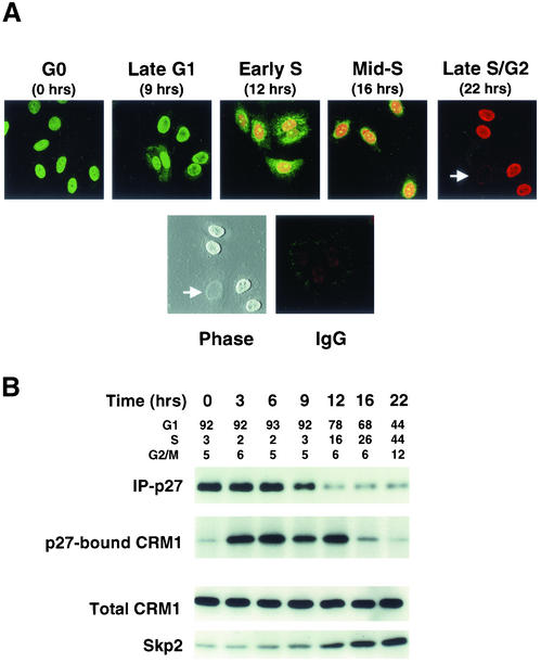
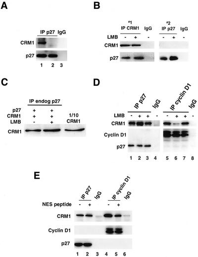
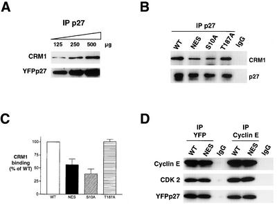
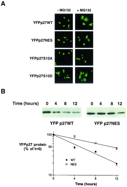
We show that p27 localization is cell cycle regulated and we suggest that active CRM1/RanGTP-mediated nuclear export of p27 may be linked to cytoplasmic p27 proteolysis in early G1. p27 is nuclear in G0 and early G1 and appears transiently in the cytoplasm at the G1/S transition. Association of p27 with the exportin CRM1 was minimal in G0 and increased markedly during G1-to-S phase progression. Proteasome inhibition in mid-G1 did not impair nuclear import of p27, but led to accumulation of p27 in the cytoplasm, suggesting that export precedes degradation for at least part of the cellular p27 pool. p27-CRM1 binding and nuclear export were inhibited by S10A mutation but not by T187A mutation. A putative nuclear export sequence in p27 is identified whose mutation reduced p27-CRM1 interaction, nuclear export, and p27 degradation. Leptomycin B (LMB) did not inhibit p27-CRM1 binding, nor did it prevent p27 export in vitro or in heterokaryon assays. Prebinding of CRM1 to the HIV-1 Rev nuclear export sequence did not inhibit p27-CRM1 interaction, suggesting that p27 binds CRM1 at a non-LMB-sensitive motif. LMB increased total cellular p27 and may do so indirectly, through effects on other p27 regulatory proteins. These data suggest a model in which p27 undergoes active, CRM1-dependent nuclear export and cytoplasmic degradation in early G1. This would permit the incremental activation of cyclin E-Cdk2 leading to cyclin E-Cdk2-mediated T187 phosphorylation and p27 proteolysis in late G1 and S phase.


Figures

Figure 1
Figure 1
p27 localization and CRM1-binding are cell cycle dependent. Cell cycle entry of quiescent MCF-7 cells was induced by the addition of 17β-estradiol at time 0. Cells were assayed at intervals thereafter for p27 localization (A) or for protein assays and cell cycle profiles (B). (A) MCF-7 cells grown on glass slides were arrested in G0 by estrogen depletion. After stimulation with estradiol, p27 levels (green) and BrdU uptake (red) were visualized by confocal fluorescence microscopy at intervals across the cell cycle. Cells negative for both p27 and BrdU staining were evident in the late S phase/G2 population (white arrow). These unstained cells are apparent on phase contrast imaging of the same field. Control cells stained with nonspecific control IgG followed by FITC and Texas Red conjugated antibodies are shown (IgG). (B) Transient binding of p27 to CRM1 occurs early in G1. p27 was immunoprecipitated at intervals across the cell cycle, p27 complexes were resolved, and immunoblots were probed for p27 and CRM1. The same cell lysates were immunoblotted for CRM1 and Skp2. The cell cycle profile at each time point was assayed by dual propidium iodide/BrdU labeling and flow cytometry.
Figure 2
Figure 2
Active nuclear export of p27. (A) Cells were grown on glass slides and synchronized as in Figure 1. Six hours after induction of cell cycle entry by 17β-estradiol addition, cells were digitonin permeabilized and subjected to export assays, fixed, and nuclear p27 visualized by indirect immunofluorescence. (B) Cells were recovered in either mid-G1 or G0 and nuclear export of p27 after digitonin permeabilization assayed as described in “Materials and Methods.” At indicated times, reactions were stopped by centrifugation and p27 was assayed in nuclei (N) and supernatant (S) fractions. p27 export was minimal after 30 min in the absence of ATP (-ATP), cytosol (-CYT), or both (-ATP/–CYT).
Figure 3
Figure 3
p27 binds CRM1 in vitro and LMB does not impair p27-CRM1 binding. (A) p27 was immunoprecipitated from cells in early G1 and incubated at 95°C for 5 min to denature heat labile-associated proteins. The supernatant containing p27 was then incubated with recombinant CRM1 together with either GTP-loaded Ran (lane 1) or GDP-loaded Ran (lane 2) for 30 min at 4°C followed by immunoprecipitation of p27. p27 antibody-bound protein A Sepharose beads did not show any nonspecific interaction with recombinant Ran or CRM1 (lane 3). (B) CRM1 was first immunoprecipitated from cells recovered in midG1 with or without prior treatment with LMB (IP #1). The supernatant was recovered and p27 was then immunoprecipitated from the CRM1-depleted lysates (IP #2). CRM1- and p27-bound proteins were immunoblotted for CRM1 and p27. Antibody only controls are shown for IP #1 and #2 (IgG). (C) The effect of LMB on binding of recombinant CRM1 to cellular p27 was assayed as in A. Equal amounts of heat stable p27 recovered from midG1 cells were reacted with recombinant RanGTP and CRM1 without (lane 1) or with (lane 2) pretreatment of the CRM1 with LMB. p27 was then immunoprecipitated and complexes were resolved and blotted for associated CRM1. One-tenth of the input recombinant CRM1 was loaded in the lane on the right. Equal amounts of p27 were immunoprecipitated in each lane (not shown). (D) Recombinant his-tagged p27 (lanes 1 through 3) or flag-tagged T286-phosphorylated cyclin D1 (lanes 5 through 7) were incubated with RanGTP and CRM1 either without (lanes 1 and 5) or with (lanes 2 and 6) pretreatment of the CRM1 with LMB. LMB was also added to the reaction mixture after complex formation (lanes 3 and 7). Antibody control lanes are also shown (lanes 4 and 8). (E) CRM1 binding assays were carried out as in D. CRM1 was preincubated with a peptide corresponding to the NES of the HIV-1 Rev protein (NES peptide) before the addition of p27 (lane 2) or cyclin D1 (lane 5).
Figure 4
Figure 4
Both LLnL and LMB increase nuclear and cytoplasmic p27 levels. (A) Mid-G1 cells were recovered at 9 h after G0 release, with or without 6 h of LLnL treatment immediately before harvesting. Lysates were immunoblotted for p27. (B) Cells were also treated as in A above and nuclear (N) and cytoplasmic (C) fractions immunoblotted for p27. Immunoblots were probed for the nuclear protein RCC1 to verify the lack of leakage of nuclear proteins into the cytoplasm. (C and D) At 6 h after G0 release, cells were incubated either with or without LMB for an additional 6 h and whole cell lysates (C) or nuclear and cytosolic fractions (D) were immunoblotted as shown. RCC1 probing verified adequacy of fractionation (not shown).
Figure 5
Figure 5
LMB does not prevent p27 nuclear export in vitro or in vivo. (A) MCF-7 cells were transfected with YFPp27WT and arrested in quiescence. Cells were digitonin permeabilized and incubated with 2.5 mg/ml cytosolic proteins and an ATP regenerating system. Incubations were carried out for the indicated times at room temperature. Where indicated, cells were pretreated with LMB or LLnL to assess the affects of these drugs on p27 nuclear export. (B) The decay of nuclear p27 fluorescence in A was visualized by direct fluorescence microscopy, photographed with a digital camera, quantitated using Carl Zeiss laser scanning software (LSM) 510, and graphed as a function of time. (C) Hela cells transfected with expression vectors for either YFPp27 or GFPp53 were fused to nontransfected NIH 3T3 cells (white arrow) in the presence or absence of LMB. The localization of either YFPp27 or GFPp53 (green) was visualized by fluorescence microscopy. Cells were fixed and stained for actin (red). Nuclei were visualized by staining the DNA with Hoechst 33258 (black and white panels). (D) p27 nuclear import was assessed by the addition of his-tagged p27 (His-p27) to digitonin-permeabilized cells in the presence of cytosolic proteins (4 μg/μl) and an ATP regenerating system. Reactions were centrifuged and nuclear (N) and supernatant (S) fractions were immunoblotted for p27. Nuclei preincubated with 200 μg/ml WGA showed no import of p27.
Figure 6
Figure 6
p27 nuclear export involves a nuclear export sequence. (A) The classical and nonclassical NES of the HIV-1Rev and EIAV Rev proteins and the sequence (amino acids 32 through 45) containing the conserved leucines in the putative p27 NES from different species are shown. (B) FITC-BSA (right panel) or FITC-BSA coupled to peptides containing the putative p27 NES (two left panels) was microinjected into the nuclei of adherant HeLa cells. Pictures at 0 and 45 min after injection were taken with identical exposure times. (C) NES mutation delays p27 export. Cells were grown on glass slides and were then transfected with YFPp27WT, YFPp27NES, YFPS10A, or YFPp27T187A. Forty-eight hours after transfection, cells were digitonin permeabilized and p27 export assayed. Nuclear export of p27 was visualized directly by photomicroscopy with a digital camera. (D) The intensity of nuclear p27 fluorescence was quantitated using Carl Zeiss Laser Scanning Software (LSM) 510 and graphed as a percentage of the maximum intensity measured at t = 0 min for each p27 allele product (p27WT, and the NES, S10A, and T187A p27 mutants).
Figure 7
Figure 7
Effects of p27 NES mutation on CRM1- and Cyclin E-binding. (A) To show the linearity of WT p27-CRM1 binding assays, increasing amounts of heat-stable YFP-p27 (125–500 μg) were incubated with fixed amounts of CRM1 and RanGTP. p27 was immunoprecipitated and immunoblots probed for associated CRM1. (B) YFPp27 was immunoprecipitated from 300 μg of lysate from cells transfected with WT, NES, S10A, or T187A YFPp27, released from protein A beads by boiling for 10 min, and incubated with recombinant CRM1 and RanGTP. p27-complexes were probed for CRM1 and p27. CRM1 binding was corrected for differences in the amount of YFPp27 expressed in the different transfectants and graphed. (C) Graphical quantification of YFP-bound CRM1. Results represent the mean ± SEM of four independent experiments. (D) MCF-7 cells were transfected with YFPp27WT or YFPp27NES expression vectors. YFP and cyclin E immunoprecipitates were assayed for associated cyclin E, Cdk 2, or p27 by immunoblotting.
Figure 8
Figure 8
Effects of p27 mutation on localization and half-life. (A) Cells were grown on glass slides and transfected with either WT, NES, S10A, or S10D YFPp27 vectors for 16 h and then treated with the proteasome inhibitor MG132 (+MG132) or without (-MG132) for an additional 8 h before fixation and photomicroscopy. (B) Cells were transfected with YFPp27WT or YFPp27NES constructs and cycloheximide (100 μg/ml) was added at 48 h posttransfection. Cells were harvested 4, 8, and 12 h after cycloheximide addition, and p27 was detected by immunoblotting with YFP-specific antibody. The decay of the p27 signal is graphed as a function of time postcycloheximide addition. Linear regression curves were fitted to calculate the half-lives of each of the mutant p27 proteins using data from repeat experiments. SE bars are shown.

References

    1. Adachi M, Fukuda M, Nishida E. Nuclear export of MAP kinase (ERK) involves a MAPK kinase (MEK)-dependent active transport mechanism. J Cell Biol. 2000;148:849–856. - PMC - PubMed
    1. Adam SA, Sterne-Marr R, Gerace L. Nuclear protein import using digitonin-permeabilized cells. Methods Enzymol. 1992;219:97–110. - PubMed
    1. Alt JR, Cleveland JL, Hannink M, Diehl JA. Phosphorylation-dependent regulation of cyclin D1 nuclear export and cyclin D1-dependent cellular transformation. Genes Dev. 2001;14:3102–3114. - PMC - PubMed
    1. Askjaer P, Bachi A, Wilm M, Bischoff FR, Weeks DL, Ogniewski V, Ohno M, Niehrs C, Kjems J, Mattaj IW, Fornerod M. RanGTP regulated interactions of CRM1 with nucleoporins and a shuttling DEADbox helicase. Mol Cell Biol. 1999;19:6276–6285. - PMC - PubMed
    1. Askjaer P, Jensen TH, Nilsson J, Englmeier L, Kjems J. The specificity of the CRM1-Rev nuclear export signal interaction is mediated by RanGTP. JBiolChem. 1998;273:33414–33422. - PubMed

Publication types

MeSH terms

LinkOut - more resources