

Adipose tissue quantification by imaging methods: a proposed classification
- PMID: 12529479
- PMCID: PMC1894646
- DOI: 10.1038/oby.2003.3
Adipose tissue quantification by imaging methods: a proposed classification
Abstract
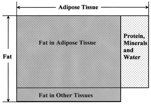
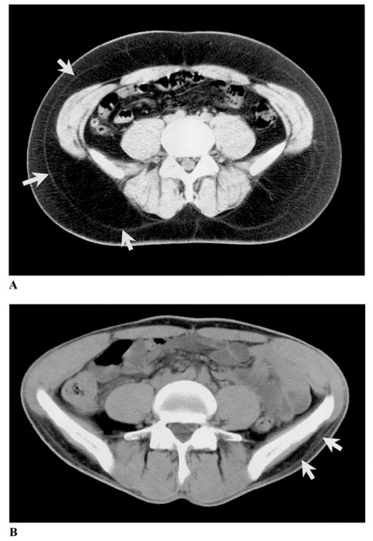
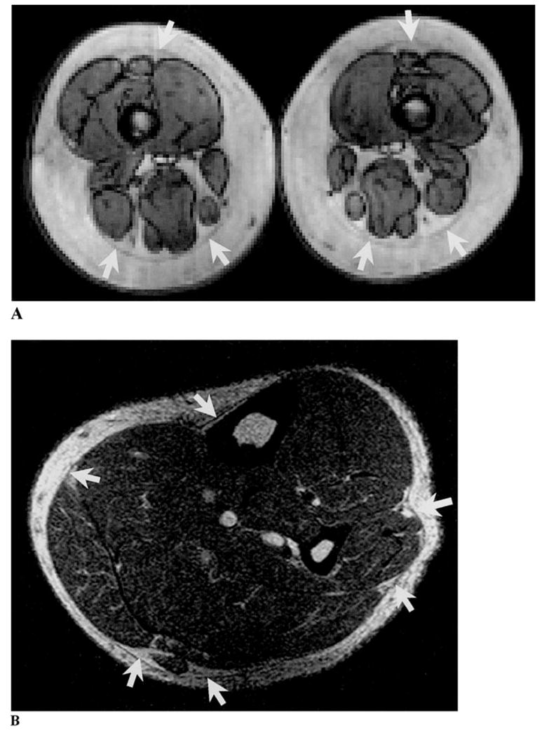
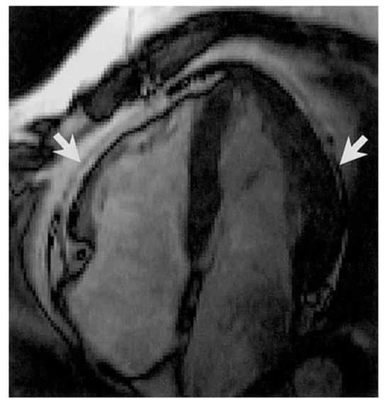
Recent advances in imaging techniques and understanding of differences in the molecular biology of adipose tissue has rendered classical anatomy obsolete, requiring a new classification of the topography of adipose tissue. Adipose tissue is one of the largest body compartments, yet a classification that defines specific adipose tissue depots based on their anatomic location and related functions is lacking. The absence of an accepted taxonomy poses problems for investigators studying adipose tissue topography and its functional correlates. The aim of this review was to critically examine the literature on imaging of whole body and regional adipose tissue and to create the first systematic classification of adipose tissue topography. Adipose tissue terminology was examined in over 100 original publications. Our analysis revealed inconsistencies in the use of specific definitions, especially for the compartment termed "visceral" adipose tissue. This analysis leads us to propose an updated classification of total body and regional adipose tissue, providing a well-defined basis for correlating imaging studies of specific adipose tissue depots with molecular processes.
Figures
References
-
- Evans DJ, Hoffmann RG, Kalkhoff RK, Kissebah AH. Relationship of body fat topography to insulin sensitivity and metabolic profiles in premenopausal women. Metabolism. 1984;33:68–75. - PubMed
-
- Vague J. The degree of masculine differentiation of obesities: a factor determining predisposition to diabetes, atherosclerosis, gout, and uric calculous disease. 1956. Nutrition. 1999;15:89–91. - PubMed
-
- Brochu M, Poehlman ET, Ades PA. Obesity, body fat distribution, and coronary artery disease. J Cardiopulm Rehabil. 2000;20:96–108. - PubMed
-
- Vanderburgh PM. Fat distribution: its physiological significance, health implications, and its adaptation to exercise training. Mil Med. 1992;157:189–92. - PubMed
Publication types
MeSH terms
Substances
Grants and funding
LinkOut - more resources
Full Text Sources
Medical
