. 2003 Jan;131(1):41-8.
doi: 10.1104/pp.014100.

Pit membrane porosity and water stress-induced cavitation in four co-existing dry rainforest tree species

Affiliations

Pit membrane porosity and water stress-induced cavitation in four co-existing dry rainforest tree species

Brendan Choat et al. Plant Physiol. 2003 Jan.

Abstract

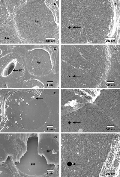
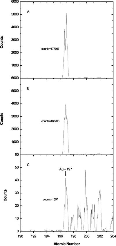
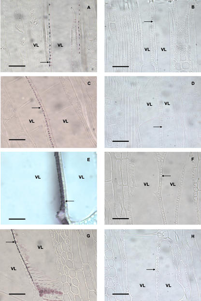
Aspects of xylem anatomy and vulnerability to water stress-induced embolism were examined in stems of two drought-deciduous species, Brachychiton australis (Schott and Endl.) A. Terracc. and Cochlospermum gillivraei Benth., and two evergreen species, Alphitonia excelsa (Fenzal) Benth. and Austromyrtus bidwillii (Benth.) Burret., growing in a seasonally dry rainforest. The deciduous species were more vulnerable to water stress-induced xylem embolism. B. australis and C. gillivraei reached a 50% loss of hydraulic conductivity at -3.17 MPa and -1.44 MPa, respectively; a 50% loss of hydraulic conductivity occurred at -5.56 MPa in A. excelsa and -5.12 MPa in A. bidwillii. To determine whether pit membrane porosity was responsible for greater vulnerability to embolism (air seeding hypothesis), pit membrane structure was examined. Expected pore sizes were calculated from vulnerability curves; however, the predicted inter-specific variation in pore sizes was not detected using scanning electron microscopy (pores were not visible to a resolution of 20 nm). Suspensions of colloidal gold particles were then perfused through branch sections. These experiments indicated that pit membrane pores were between 5 and 20 nm in diameter in all four species. The results may be explained by three possibilities: (a) the pores of the expected size range were not present, (b) larger pores, within the size range to cause air seeding, were present but were rare enough to avoid detection, or (c) pore sizes in the expected range only develop while the membrane is under mechanical stress (during air seeding) due to stretching/flexing.


Figures

Figure 1
Figure 1
Vulnerability curves in species of contrasting leaf phenology showing PLC versus Ψx in excised air-dried stem segments. Each point represents PLC for single branch. Vulnerability curves were obtained by fitting the exponential sigmoidal equation PLC = 100/(1 + exp{axb]}), to the data, where a describes the slope of the curve and b is the Ψx at which a PLC of 50 occurs (Pammenter and Vander Willigen, 1998). Values for a, b, and r2 are given in Table I.
Figure 2
Figure 2
Scanning electron micrographs of inter-vessel pit membranes in each of the species. Each picture shows a longitudinal section of xylem tissue in which one secondary wall has been partially or fully removed to reveal pit membranes. Micrographs in the right column show higher resolution images in which the cellulose microfibrillar structure of membranes is clearly visible. Black dots shown in the right column represent circular pores of the size calculated for each species in Table III. A, Intact membranes (PM) of A. bidwillii with cellulose microfibrils visible (×35,000). The surrounding wall has been impregnated with lignin (LW), and microfibrils cannot be discerned. B, Closer view (×50,000) of pit membrane in A. bidwillii. C, Intact membrane (PM) and torn membrane revealing underlying pit channel (PC) in A. excelsa (×10,000). D, Detail of pit membrane in A. excelsa (×35,000) with margin of membrane visible to right side of micrograph. E, Pit membranes of B. australis (×11,000). Overlapping layers of microfibrils are visible at the top of the membrane in the center of the micrograph (arrow). F, Detail of membrane in B. australis showing closer view of overlapping primary wall layers (×35,000). G, Pit membrane (PM) of C. gillivraei with overarching secondary walls (SW; ×11,000). H, Closer view of pit membrane in C. gillivraei at (×35,000). Margin of membrane and secondary wall is visible on right side of micrograph.
Figure 3
Figure 3
Detection of gold (Au) in perfusate using ICP-MS. Graphs show integrated counts of gold for concentration standard (A; 5 μL L−1 gold colloid in suspension), representative sample of 5-nm gold colloid perfusate from C. gillivraei (B), and representative sample of 20-nm gold colloid perfusate from C. gillivraei (C). Note the scale of y axis for 20-nm sample differs from 5-nm sample and concentration standard.
Figure 4
Figure 4
Light micrographs show tangential longitudinal sections through the xylem tissue of each species after perfusing branch segments with gold colloid suspensions A. bidwillii (A and B), A. excelsa (C and D), B. australis (E and F), and C. gillivraei (G and H). All micrographs show two adjacent xylem vessels, with vessel lumens labeled VL. Pictures in the left column show branches that have been perfused with suspensions containing 20-nm gold colloids (A, C, E, and G). Gold particles have accumulated at inter-vessel pit membranes (arrows) and in pit cavities of each species. Stem sections shown in the right column have been perfused with suspension containing 5-nm gold colloids (B, D, F, and H); pit membranes and pit cavities show little accumulation of gold particles. Scale bars = 50 μm.

References

    1. Alder NN, Sperry JS, Pockman WT. Root and stem xylem embolism, stomatal conductance, and leaf turgor in Acer grandidentatum populations along a soil moisture gradient. Oecologia. 1996;105:293–301. - PubMed
    1. Butterfield BG, Meylan BA. Cell wall hydrolysis in the tracheary elements of the secondary xylem. In: Baas P, editor. New Perspectives in Wood Anatomy. The Hague, The Netherlands: Junk Publishers; 1982.
    1. Cochard H, Cruiziat P, Tyree MT. Use of positive pressures to establish vulnerability curves: further support for the air-seeding hypothesis and implications for pressure-volume analysis. Plant Physiol. 1992;100:205–209. - PMC - PubMed
    1. Crombie DS, Hipkins MF, Milburn JA. Gas penetration of pit membranes in the xylem of Rhododendron as the cause of acoustically detected sap cavitation. Aust J Plant Physiol. 1985;12:445–453.
    1. Cronshaw J. The fine structure of the pits of Eucalyptus regnans (F. Muell.) and their relation to the movement of liquids into wood. Aust J Bot. 1960;8:51–57.

LinkOut - more resources