. 2003 Jan;131(1):93-101.
doi: 10.1104/pp.011585.

Elicitor activity of a fungal endopolygalacturonase in tobacco requires a functional catalytic site and cell wall localization

Affiliations

Elicitor activity of a fungal endopolygalacturonase in tobacco requires a functional catalytic site and cell wall localization

Georges Boudart et al. Plant Physiol. 2003 Jan.

Abstract

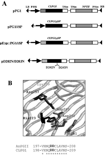
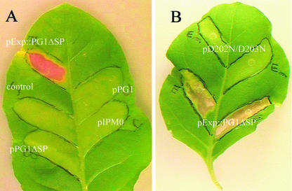
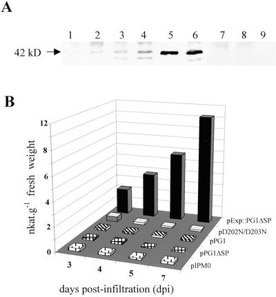
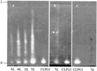
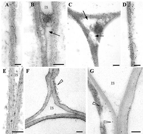
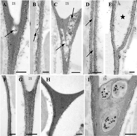
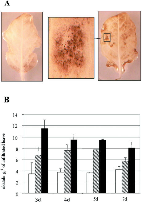
CLPG1, an endopolygalacturonase (endoPG) gene of Colletotrichum lindemuthianum, was transferred to tobacco (Nicotiana tabacum) leaves by using the Agrobacterium tumefaciens transient delivery system. The following four constructs were prepared: CLPG1, with or without its signal peptide (SP; PG1, PG1deltaSP); CLPG1 with the tobacco expansin1 SP instead of its own SP (Exp::PG1deltaSP); and a mutated version of the latter on two amino acids potentially involved in the catalytic site of CLPG1 (D202N/D203N). Chlorotic and necrotic lesions appeared 5 to 7 d postinfiltration, exclusively in response to CLPG1 fused to the expansin SP. The lesions were correlated to the production of an active enzyme. Necrosis-inducing activity, as well as endoPG activity, were completely abolished by site-directed mutagenesis. Ultrastructural immunocytolocalization experiments indicated that the expansin SP addressed CLPG1 to the cell wall. Staining of parenchyma cells revealed the progressive degradation of pectic material in junction zones and middle lamella as a function of time after infiltration, ultimately leading to cell separation. A 30% decrease in the GalUA content of the cell walls was simultaneously recorded, thereby confirming the hydrolytic effect of CLPG1 on pectic polysaccharides, in planta. The elicitor activity of CLPG1 was further illustrated by the induction of defense responses comprising active oxygen species and beta-1,3-glucanase activity, before leaf necrosis. Altogether, the data demonstrate that an appropriate SP and a functional catalytic site are required for the proper expression and elicitor activity of the fungal endoPG CLPG1 in tobacco.


Figures

Figure 1
Figure 1
Measurement of PGIP activity against CLPG1 in protein extracts from tobacco cv Samsun NN, Arabidopsis (ecotype Columbia), and French bean seedlings. The experiment was repeated two times with the same results.
Figure 2
Figure 2
A, Structure of T-DNA constructs transferred to plant cells via A. tumefaciens. Theed into the pIPM0 binary vector with (pPG1) or without (pPG1ΔSP) its own SP, or with the SP of the tobacco expansin1 gene (pExp::PG1ΔSP). An additional construct was obtained from pExp::PG1ΔSP by site-directed mutagenesis of Asp-202 and Asp-203 codons putatively involved in the catalytic site of the enzyme, giving rise to two Asn (pD202N/D203N). Each construct was under the control of cauliflower mosaic virus 35S promoter and of the nopaline synthase terminator (tNos). B, Three-dimensional representation of the putative catalytic site of CLPG1, as modelized by analogy with AnPGII endoPG of Aspergillus niger (van Santen et al., 1999) using the SWISS-MODEL software (http://swissmodel.expasy.org/; Guex and Peitsch, 1999). The amino acid sequences of the catalytic site of AnPGII endoPG (upper line) and of CLPG1 (lower line) are represented (amino acid identity shown by asterisks).
Figure 3
Figure 3
Symptoms induced in tobacco leaves infiltrated with A. tumefaciens carrying: A, pPG1, pPG1ΔSP, pExp::PG1ΔSP, or the empty vector; and B, pExp::PG1ΔSP or pD202N/D203N. Leaves were also infiltrated with infiltration medium (10 mm MgSO4 + 250 μm acetosyringone) as a control. The infiltration area was outlined with a black marker pen. Leaves were photographed 2 weeks postinfiltration, using a CCD-IRIS color video camera. Necrosis was only observed upon A. tumefaciens transformation with pExp::PG1ΔSP.
Figure 4
Figure 4
Time course measurement of endoPG in protein extracts prepared from whole leaves infiltrated with agrobacteria carrying either pPG1, pPG1ΔSP, pExp::PG1ΔSP, or the mutant pD202N/D203N. A, Western-blot analysis of protein extracts of tobacco leaves agro-infiltrated with pExp::PG1ΔSP (lanes 1–4), pD202N/D203N (lane 6), pPG1 (lane 7), pPG1ΔSP (lane 8), or pIPM0 (lane 9). Lanes 1 through 4 corresponded to 3, 4, 5, and 7 dpi, respectively, and lanes 6 through 9 to 7 dpi. Pure CLPG1 (42 kD) was in lane 5. Proteins were separated by SDS-PAGE on a 10% (w/v) polyacrylamide gel and blotted onto a nitrocellulose membrane. Proteins cross-reacting with a CLPG1 polyclonal antiserum were revealed with a secondary goat anti-rabbit antiserum conjugated to alkaline phosphatase. Alkaline phosphatase activity was revealed with nitroblue tetrazolium and 5-bromo-4-chloro-3-indolyl phosphate. B, endoPG activity was colorimetrically assessed. The experiment was repeated three times with the same time course increase of endoPG activity upon infiltration with pExp::CLPG1ΔSP.
Figure 5
Figure 5
Zymogram of endoPG activity in protein extracts recovered from tobacco leaves 3 to 7 dpi with A. tumefaciens carrying pExp::PG1ΔSP (A) and 7 dpi with A. tumefaciens carrying pD202N/D203N (C). endoPG activity was assayed by analytical isoelectric focusing. The active endoPG focused at a pI identical to pure CLPG1 protein (pI = 10.1). B, An additional endoPG isoform focusing at a neutral or slightly acidic pI value (arrow) was also revealed.
Figure 6
Figure 6
Immunogold labeling of leaf parenchyma cell walls 4 (A, B, and E) and 7 (C and D) d postinfiltration with A. tumefaciens carrying pExp::PG1ΔSP (A–D) or the mutant pD202N/D203N (E). Labeling was achieved with antiserum against CLPG1 and gold-conjugated goat antiserum to rabbit IgG. Gold particles were found within the cell walls (A and B) and in intercellular spaces (C and D) at nearly identical levels at 4 and 7 dpi. Note degradation of pectic material in intercellular spaces (IS; arrows). Gold particles were also observed within the cell walls and intercellular spaces 4 dpi with A. tumefaciens carrying the mutant pD202N/D203N (E), without detectable pectin degradation. A few gold particles were observed in leaves agro-infiltrated with the empty vector (cell wall-cytoplasm interface, arrowhead, F), and in sections treated with the secondary antibody alone (arrowheads, G). Sections were contrasted with uranyl acetate. Scale bar = 0.3 μm.
Figure 7
Figure 7
Electron micrographs of parenchyma cell walls stained with the PATAg reagent for polysaccharides visualization. A through E, Plant expressing Exp::PG1ΔSP: A, 4 dpi, the solubilization of cell wall components was limited to the cell corners (arrow); B, was also visible along the cell walls between two tricellular junctions; C, 7 dpi, cell wall degradation was clearly visible in the cell corner where large areas were cleared out (arrow); and D, in the middle lamella (arrow), ultimately leading to separation of the two walls (E). F and G, Control, non-agro-infiltrated tissues. H, Plants expressing D202N/D203N were heavily stained with no apparent alteration of the walls. I, Three bacteria in the corner of an intercellular space (IS). Note the absence of cell wall degradation in contact to bacteria. Bar = 0.3 μm.
Figure 8
Figure 8
AOS detection and time course measurement of β-1,3-glucanase activity in agro-infiltrated tobacco leaves. A, AOS were detected as dark-brown deposits (right, insert) 5 dpi in leaf tissues expressing Exp::PG1ΔSP using the DAB uptake method. AOS were not detected in leaves agro-infiltrated with the empty vector (left). B, Time course measurement of β-1,3-glucanase activity of tobacco leaves infiltrated with agrobacteria carrying either pExp::PG1ΔSP (black box) or the empty vector (hatched box). Control leaves (white box) were infiltrated with infiltration medium (10 mm MgSO4 + 250 μm acetosyringone). β-1,3-glucanase activity was colorimetrically assessed.

References

    1. Armand S, Wagemaker MJ, Sanchez-Torres P, Kester HC, van Santen Y, Dijkstra BW, Visser J, Benen JA. The active site topology of Aspergillus niger endopolygalacturonase II as studied by site-directed mutagenesis. J Biol Chem. 2000;275:691–696. - PubMed
    1. Atkinson RG, Schröder R, Hallett IC, Cohen D, MacRae EA. Overexpression of polygalacturonase in transgenic apple trees leads to a range of novel phenotypes involving changes in cell adhesion. Plant Physiol. 2002;129:122–133. - PMC - PubMed
    1. Bendahmane A, Querci M, Kanyuka K, Baulcombe DC. Agrobacterium transient expression system as a tool for the isolation of disease resistance genes: application to the Rx2 locus in potato. Plant J. 2000;21:73–81. - PubMed
    1. Blumenkrantz N, Asboe-Hansen G. A new method for quantitative determination of uronic acids. Anal Biochem. 1973;54:484–489. - PubMed
    1. Boudart G, Lafitte C, Barthe JP, Frasez D, Esquerré-Tugayé MT. Differential elicitation of defense responses by pectic fragments in bean seedlings. Planta. 1998;206:86–94.

MeSH terms

LinkOut - more resources