Comment
. 2003 Jan;111(2):231-9.
doi: 10.1172/JCI16672.

Hydroxyurea induces fetal hemoglobin by the nitric oxide-dependent activation of soluble guanylyl cyclase

Affiliations
Comment

Hydroxyurea induces fetal hemoglobin by the nitric oxide-dependent activation of soluble guanylyl cyclase

Vladan P Cokic et al. J Clin Invest. 2003 Jan.

Abstract

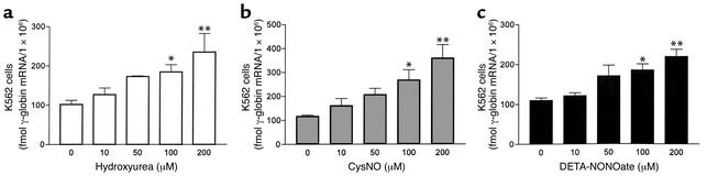
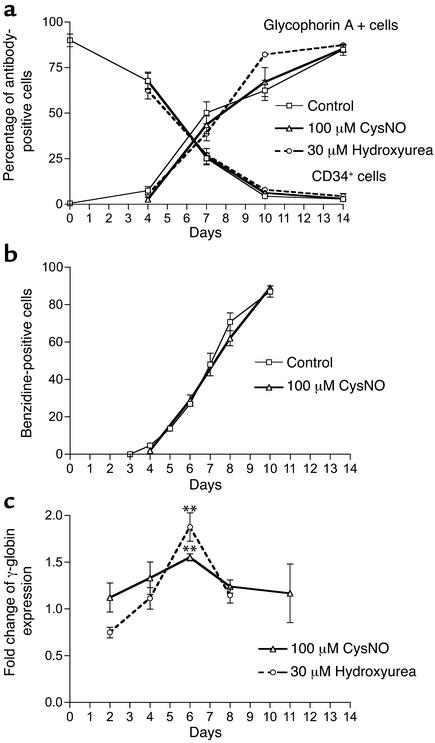
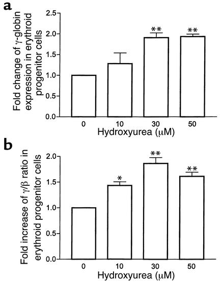
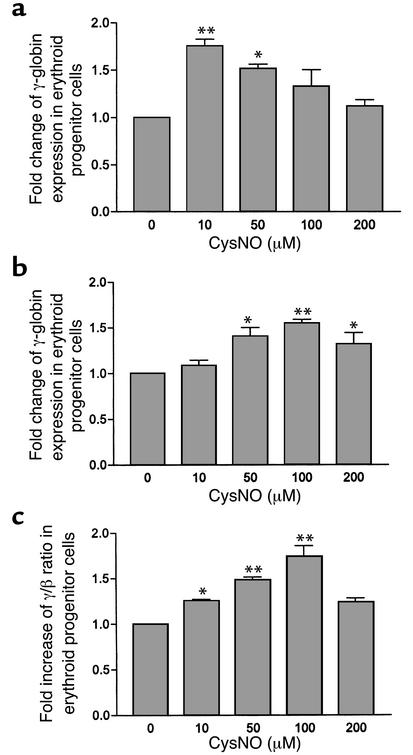
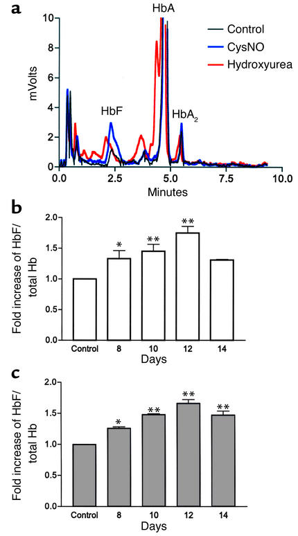
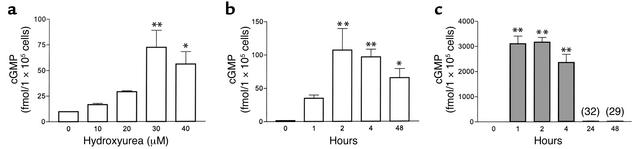
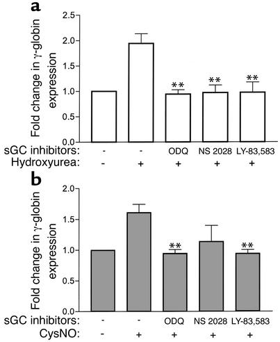
Hydroxyurea treatment of patients with sickle-cell disease increases fetal hemoglobin (HbF), which reduces hemoglobin S polymerization and clinical complications. Despite its use in the treatment of myeloproliferative diseases for over 30 years, its mechanism of action remains uncertain. Recent studies have demonstrated that hydroxyurea generates the nitric oxide (NO) radical in vivo, and we therefore hypothesized that NO-donor properties might determine the hemoglobin phenotype. We treated both K562 erythroleukemic cells and human erythroid progenitor cells with S-nitrosocysteine (CysNO), an NO donor, and found similar dose- and time-dependent induction of gamma-globin mRNA and HbF protein as we observed with hydroxyurea. Both hydroxyurea and CysNO increased cGMP levels, and the guanylyl cyclase inhibitors ODQ, NS 2028, and LY 83,538 abolished both the hydroxyurea- and CysNO-induced gamma-globin expression. These data provide strong evidence for an NO-derived mechanism for HbF induction by hydroxyurea and suggest possibilities for therapies based on NO-releasing or -potentiating agents.


Figures

Figure 1
Figure 1
Hydroxyurea and NO induce γ-globin gene expression in K562 cells. (ac) Hydroxyurea (white bars), CysNO (gray bars), and DETA-NONOate (black bars) induce γ-globin gene expression in K562 cells measured as femtomoles of γ-globin mRNA per 106 cells after 24 hours of treatment. Values represent means ± SEM (n = 3). *P < 0.05 and **P < 0.01 versus untreated cells (0).
Figure 2
Figure 2
Flow cytometry of erythroid cells during their differentiation. (a) We used glycophorin A and CD34 antibodies to detect changes in the percentage of antibody-positive cells during erythroid differentiation of CD34+ cells. Hydroxyurea and CysNO did not influence erythroid differentiation (n = 5 or 6). (b) After 24 hours of incubation with 100 μM CysNO at different time points of erythroid differentiation, the benzidine-positive cells were scored and expressed as a percentage of the number of total cells (n = 3). (c) At different time points during erythroid differentiation, after 24 hours of incubation with hydroxyurea or CysNO, we harvested erythroid cells (corresponding to values for days in the figure) and compared induction of γ-globin mRNA expression. Data are normalized by cell number and, for each culture experiment, are expressed as ratios to values for controls not treated with hydroxyurea or CysNO. Values represent means ± SEM (n = 3 or 4). **P < 0.01 versus control.
Figure 3
Figure 3
Hydroxyurea induces γ-globin gene expression in erythroid progenitor cells. (a and b) Hydroxyurea induces γ-globin gene expression (a) and increases the γ/β ratio (b) in erythroid progenitor cells. Erythroid progenitor cells were treated with hydroxyurea on day 4, and after 48 hours of incubation total RNA was iso-lated. Data are normalized by cell number and, for each culture experiment, are expressed as ratios to values for controls not treated with hydroxyurea. Values represent means ± SEM (n = 4). *P < 0.05 and **P < 0.01 versus untreated cells (0).
Figure 4
Figure 4
CysNO induces γ-globin gene expression in erythroid progenitor cells. (a) We treated erythroid progenitor cells with CysNO on day 3, and after 24 hours of incubation we harvested them and measured γ-globin expression (n = 3). (b and c) On day 5 of erythroid cell culture, we added CysNO, and we measured mRNA levels on day 6; CysNO increases γ-globin expression (normalized as in Figure 3) (b) as well as the γ/β ratio (c). Values represent means ± SEM (n = 4). *P < 0.05 and **P < 0.01 versus untreated cells (0).
Figure 5
Figure 5
HPLC analysis of changes in HbF during maturation of erythroid cells. On day 4 of erythroid cell culture, we treated erythroid progenitor cells either with hydroxyurea (white bars) or CysNO (gray bars) and measured HbF versus total hemoglobin levels at different time points during erythroid maturation. (a) Chromatograms of HPLC analyses of hemoglobins produced by erythroid precursor cells during treatment with hydroxyurea (30 μM, red line) and CysNO (100 μM, blue line) on day 10. (b and c) Increases in the percent of HbF to total hemoglobin after treatment with 30 μM hydroxyurea (b) and 100 μM CysNO (c). Values represent means ± SEM (n = 4). *P < 0.05 and **P < 0.01 versus control.
Figure 6
Figure 6
Effects of hydroxyurea and CysNO on intracellular cGMP levels in erythroid progenitor cells. (a) Hydroxyurea (white bars) was added on day 4 to a suspension of erythroid progenitor cells, and intracellular cGMP levels were measured after 48 hours. (b) Hydroxyurea (30 μM) was added on day 4, and cGMP levels were measured within the first 4 hours, as well as after 48 hours. (c) CysNO (100 μM, gray bars) was added on day 4, with measurements taken for the first 4 hours as well as after 24 and 48 hours. Values represent means ± SEM (n = 3). *P < 0.05 and **P < 0.01 versus untreated cells (0).
Figure 7
Figure 7
Inhibition of γ-globin induction by sGC inhibitors. (a) Before incubation with hydroxyurea (30 μM), erythroid progenitor cells were pretreated with the sGC inhibitors ODQ (10 μM), NS 2028 (0.5 μM), and LY 83,583 (5 μM) for 30 minutes, 15 minutes, and 12 hours, respectively. Hydroxyurea (white bars) was added on day 4, and γ-globin mRNA levels were measured after 48 hours. (b) After preincubation with sGC inhibitors (as for preincubation for hydroxyurea), erythroid progenitor cells were treated with CysNO (100 μM, gray bars) on day 5, and γ-globin mRNA levels were measured after 24 hours. Values represent means ± SEM (n = 3). **P < 0.01 versus cells treated with hydroxyurea or CysNO.

Comment on

References

    1. Charache S, et al. Effect of hydroxyurea on the frequency of painful crises in sickle cell anemia. Investigators of the Multicenter Study of Hydroxyurea in Sickle Cell Anemia. N. Engl. J. Med. 1995;332:1317–1322. - PubMed
    1. Charache S. Mechanism of action of hydroxyurea in the management of sickle cell anemia in adults. Semin. Hematol. 1997;34:15–21. - PubMed
    1. Atweh GF, Schechter AN. Pharmacologic induction of fetal hemoglobin: raising the therapeutic bar in sickle cell disease. Curr. Opin. Hematol. 2001;8:123–130. - PubMed
    1. Letvin NL, et al. Influence of cell cycle phase-specific agents on simian fetal hemoglobin synthesis. J. Clin. Invest. 1985;75:1999–2005. - PMC - PubMed
    1. Platt OS, et al. Hydroxyurea enhances fetal hemoglobin production in sickle cell anemia. J. Clin. Invest. 1984;74:652–656. - PMC - PubMed