. 2003 Jan;111(2):265-73.
doi: 10.1172/JCI15109.

A methylated oligonucleotide inhibits IGF2 expression and enhances survival in a model of hepatocellular carcinoma

Affiliations

A methylated oligonucleotide inhibits IGF2 expression and enhances survival in a model of hepatocellular carcinoma

Xiaoming Yao et al. J Clin Invest. 2003 Jan.

Abstract

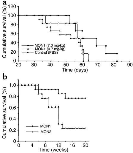
IGF-II is a mitogenic peptide that has been implicated in hepatocellular oncogenesis. Since the silencing of gene expression is frequently associated with cytosine methylation at cytosine-guanine (CpG) dinucleotides, we designed a methylated oligonucleotide (MON1) complementary to a region encompassing IGF2 promoter P4 in an attempt to induce DNA methylation at that locus and diminish IGF2 mRNA levels. MON1 specifically inhibited IGF2 mRNA accumulation in vitro, whereas an oligonucleotide (ON1) with the same sequence but with nonmethylated cytosines had no effect on IGF2 mRNA abundance. MON1 treatment led to the specific induction of de novo DNA methylation in the region of IGF2 promoter hP4. Cells from a human hepatocellular carcinoma (HCC) cell line, Hep 3B, were implanted into the livers of nude mice, resulting in the growth of large tumors. Animals treated with MON1 had markedly prolonged survival as compared with those animals treated with saline or a truncated methylated oligonucleotide that did not alter IGF2 mRNA levels in vitro. This study demonstrates that a methylated sense oligonucleotide can be used to induce epigenetic changes in the IGF2 gene and that inhibition of IGF2 mRNA accumulation may lead to enhanced survival in a model of HCC.


Figures

Figure 1
Figure 1
IGF2 expression in human HSK 09 cells treated with the methylated oligonucleotide MON1, as assessed by RT-PCR. IGF2 mRNA abundance was normalized by comparing its density with that of either IGF2 genomic DNA or an internal control, β-actin.
Figure 2
Figure 2
Inhibition of IGF2 expression by MON1 oligonucleotide in liver cancer cells. (a) Hep 3B and Hep G2 cells were seeded in six-well plates and were treated with MON1 (2 μM) in a low-serum medium. As described in Methods, both IGF2 DNA and IGF2 mRNA were coamplified by the same primer set that encompasses intron 8 of the human IGF2 gene. (b) Hep 3B cells were treated with varying doses (0.0–10.0 μM) of MON1 for 60 hours, and IGF2 mRNA abundance was quantitated by RT-PCR. (c) Northern blot analysis of IGF2 mRNA in Hep 3B cells after MON1 treatment using a probe covering IGF2 exons 7, 8, and 9.
Figure 3
Figure 3
Specific inhibition of IGF2 expression with the methylated 22-mer oligonucleotide MON1. (a) Hep 3B cells were treated with the methylated 22-mer MON1 and the truncated methylated 19-mer MON2. The abundance of IGF2 mRNA was quantitated by RT-PCR in duplicate samples. Controls were incubated with PBS alone. (b) Hep 3B cells were treated with methylated and unmethylated oligonucleotides. Lane 1 shows a 100-bp marker; lane 2, PBS control; lane 3, unmethylated antisense of 22-mer ON1; lane 4, unmethylated 22-mer ON1; lane 5, methylated MON1; lane 6, unmethylated 19-mer ON2; and lane 7, methylated 19-mer MON2.
Figure 4
Figure 4
Nucleic enrichment of MON1 in Hep 3B tumor cells. MON1 was labeled using a fluorescence kit and was incubated with Hep 3B cells in the liposome-encapsulated form. After 24 hours of incubation, tumor cells were washed with PBS and examined by confocal microscopy. The arrows point to nuclei concentrating the fluorescent MON1 (oil, ×400). Images were acquired from two separate fields with FITC (left) and combined (right) settings, respectively.
Figure 5
Figure 5
DNA methylation analysis of IGF2 by Southern blotting. (a) Location of HpaII (CCGG) sites and labeling probe. (b) Southern blotting. Lane 1 shows a 1-kb marker; lanes 2–4, PBS control DNA; and lanes 5–7, MON1-treated DNA. Genomic DNA was digested with PvuII and then either with HpaII sensitive to DNA methylation or by MspI insensitive to DNA methylation. MON1 induced DNA methylation at the promoter site of IGF2, and thus the 1084-bp band was resistant to DNA methylation–sensitive HpaII (lanes 5 and 6) but not to DNA methylation–insensitive MspI (lane 7). DNA from control cells (lanes 3 and 4) was completely digested by HpaII, indicating that control DNA was not methylated in that region.
Figure 6
Figure 6
DNA methylation at specific CpG dinucleotides in the hP4 region as measured by bisulfite sequencing. (a) Scheme of the human IGF2 promoters (hP1–hP4, indicated by arrows) and CpG sites (in bold italics) in hP4. The locations of MON1-targeted sequence are underlined, and the putative TATA box is enclosed in the rectangle. The coding region of IGF2 is depicted in black boxes (GenBank accession no. C006408, 53861–53677). E1–E9 refers to exons 1–9. (b) DNA methylation profile in the hP4 region. Genomic DNA was treated with sodium bisulfite. The bottom strand was amplified with PCR and then sequenced using a sequencing kit. CpGs (5 and 6) were within the MON1 sequence. Note that almost all cytosines in hP4 are unmethylated in control cells and thus were converted into thymidines after treatment. The preservation of the C residues indicates that they were methylated, demonstrating that MON1 treatment induces DNA methylation in the target region.
Figure 7
Figure 7
Cumulative survival rate of nude mice implanted with Hep 3B tumor cells. (a) In study 1, athymic nude mice were implanted with Hep 3B tumor cells (107 cells). Ten days after tumor cell implantation, animals were randomly assigned into treatment groups and began to receive MON1 at doses of 0.7 mg/kg and 7 mg/kg and PBS control. Animals were dosed intravenously through the tail vein twice a week. At the end of the study, animals receiving MON1 treatment (7 mg/kg) had significantly prolonged survival in a clear dose-dependent manner (P < 0.05 by the Mantel-Cox log-rank test).(b) In study 2, nude mice were implanted with 107 Hep 3B cells. Four weeks after implantation, animals were randomly assigned to receive MON2 (10 mg/kg, n = 13) or MON1 (10 mg/kg, n = 13). All treatments were administered through tail-vein injection twice a week. The Mantel-Cox log-rank test shows a significantly prolonged survival in animals receiving MON1 treatment (10 mg/kg) as compared with those receiving MON2 treatment (P < 0.05).

References

    1. Nakae J, Kido Y, Accili D. Distinct and overlapping functions of insulin and IGF-I receptors. Endocr. Rev. 2001;22:818–835. - PubMed
    1. Cariani E, et al. Differential expression of insulin-like growth factor II mRNA in human primary liver cancers, benign liver tumors, and liver cirrhosis. Cancer Res. 1988;48:6844–6849. - PubMed
    1. Kim SO, Park JG, Lee YI. Increased expression of the insulin-like growth factor I (IGF-I) receptor gene in hepatocellular carcinoma cell lines: implications of IGF-I receptor gene activation by hepatitis B virus X gene product. Cancer Res. 1996;56:3831–3836. - PubMed
    1. Pulford DJ, Falls JG, Killian JK, Jirtle RL. Polymorphisms, genomic imprinting and cancer susceptibility. Mutat. Res. 1999;436:59–67. - PubMed
    1. Zhang L, et al. Gene expression profiles in normal and cancer cells. Science. 1997;276:1268–1272. - PubMed

Publication types

MeSH terms