. 2003 Jan;111(2):275-83.
doi: 10.1172/JCI16530.

Prolactin modulates the naive B cell repertoire

Affiliations

Prolactin modulates the naive B cell repertoire

Elena Peeva et al. J Clin Invest. 2003 Jan.

Abstract

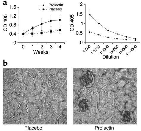
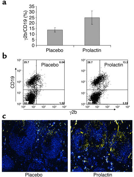
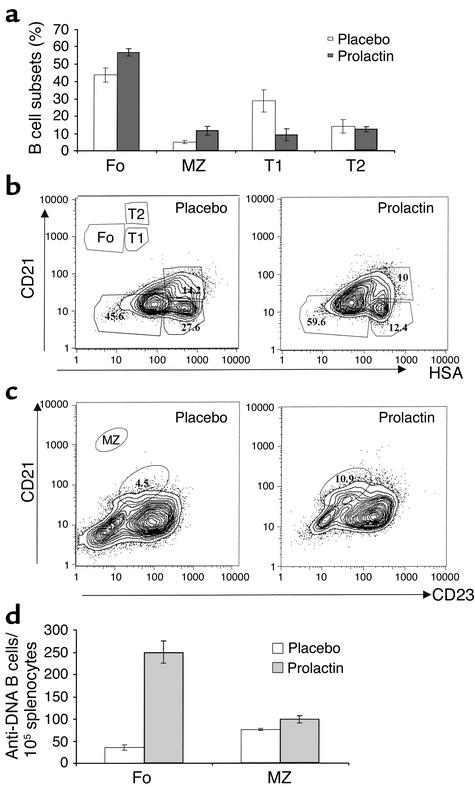
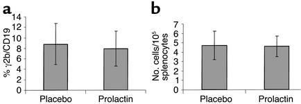
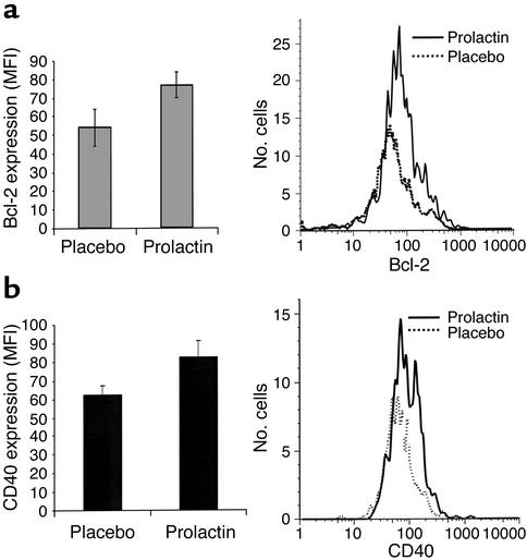
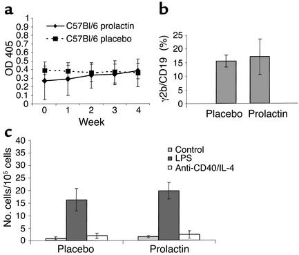
Prolactin is a peptide hormone produced by the anterior pituitary gland that is critical in lactation. Prolactin can also be produced by lymphocytes, and both B and T cells express prolactin receptors. These findings have suggested that prolactin has immunomodulatory functions. Studies in spontaneously autoimmune hosts have demonstrated a role for prolactin in augmenting autoreactivity. We chose to analyze prolactin effects on anti-DNA B cells in nonspontaneously autoimmune female BALB/c mice transgenic for the heavy chain of an anti-DNA antibody. Treatment with prolactin for 4 weeks induced a lupus-like phenotype with an increased number of transgene-expressing B cells, elevated serum anti-DNA antibody titers, and glomerular immunoglobulin deposits. Prolactin caused a decrease in the population of transitional B cells and an increase in mature follicular and marginal zone B cells. The DNA-reactive B cells had a follicular cell phenotype. Anti-DNA hybridomas demonstrated that prolactin alters selection of the naive B cell repertoire. The expansion and activation of anti-DNA B cells in prolactin-treated R4A-gamma2b BALB/c mice was dependent on the presence of CD4(+) T cells. Finally, treatment with prolactin was unable to break tolerance in R4A-gamma2b transgenic C57Bl/6 mice, suggesting that responsiveness of the immune system to prolactin is genetically determined.


Figures

Figure 1
Figure 1
(a) Serum titers of anti-dsDNA antibodies. Fourteen R4A-γ2b BALB/c mice were injected subcutaneously with 100 μg of murine prolactin daily and 12 were injected with placebo daily for 4 weeks. Sera collected before initiation of treatment and weekly thereafter showed an increase in anti-DNA titer in placebo-treated mice. Anti-DNA reactivity assayed from serum obtained at the end of the treatment showed that prolactin-treated mice had significantly higher titers of anti-DNA antibody than placebo-treated mice (P = 0.023). OD 405, optical density at 405 nanometers. (b) Immunohistochemistry of kidney sections. Fourteen prolactin-treated and 12 placebo-treated R4A-γ2b BALB/c mice were sacrificed after 4 weeks of treatment. The kidneys were stained for IgG deposition. Anti-DNA deposits were found in prolactin-treated mice but not in placebo-treated mice. Representative sections from mice treated with prolactin or placebo are shown. Magnification, ×40.
Figure 2
Figure 2
Analysis of peripheral B cell lymphocytes. Splenocytes were isolated from seven murine prolactin–treated and seven placebo-treated mice after 4 weeks of treatment. (a) A higher percentage of B cells express γ2b in prolactin-treated than in placebo-treated mice (P = 0.01). The total number of γ2b cells in the spleens of prolactin-treated mice was 3.98 ± 0.84 × 10⁶, as compared with 2.3 ± 0.53 × 10⁶ in placebo-treated mice. (b) γ2b staining of splenocytes Representative dot plot showing an increased number of γ2b-expressing cells in prolactin-treated mice. (c) Localization of IgG2b-expressing B cells. B cell regions in the spleen were identified with anti-IgM staining (blue); γ2b-expressing B cells were labeled with anti-γ2b (yellow). The spleens of prolactin-treated mice displayed an expansion of the γ2b B cells, which were found mainly in the follicles. In spleens of placebo-treated mice, transgene-bearing B cells were localized to the T/B cell interface.
Figure 3
Figure 3
B cell maturation. (a) Splenocytes from nontransgenic BALB/c mice treated with murine prolactin (n = 5) or placebo (n = 3) were stained for CD19, CD21, CD23, and HSA and were analyzed for T1, T2, marginal zone, and follicular subsets. B cell subsets were analyzed on the basis of data obtained with CD21 and HSA staining for the T1, T2, and follicular subsets and CD21 and CD23 staining for the marginal zone subset. (b) In prolactin-treated mice, the numbers of immature HSAhigh transitional B cells were reduced (P = 0.002). In the mature HSAlow B cell population, the numbers of follicular (CD21intermed/HSAlow) B cells were increased (P = 0.002). (c) The marginal zone B cell subset (CD21high/CD23low) was increased in prolactin-treated mice (P = 0.005) (d) ELISpot assay of follicular and marginal zone B cells pooled from four ovine prolactin–treated or four placebo-treated mice demonstrated an increase in the number of spontaneously secreting DNA-reactive follicular B cells. MZ, marginal zone; Fo, follicula.
Figure 4
Figure 4
Anti-DNA B cells in CD4 knockout R4A-γ2b transgenic BALB/c mice. Splenocytes were isolated from seven murine prolactin–treated and seven placebo-treated CD4 knockout R4A-γ2b BALB/c mice after 4 weeks of treatment and were evaluated for γ2b expression and the number of spontaneously activated DNA-reactive B cells. (a) Prolactin- and placebo-treated mice displayed the same difference in the percentage of B cells expressing γ2b. The total numbers of γ2b cells in the spleens of prolactin- and placebo-treated mice were 1.82 ± 0.76 × 10⁶ and 1.67 ± 0.51 × 10⁶, respectively. (b) ELISpot assay showed that prolactin did not induce an increase in the number of anti-DNA antibody–secreting B cells (cells).
Figure 5
Figure 5
Gene expression. B cells from ovine prolactin– and placebo-treated mice were analyzed for cell surface and intracellular markers. (a) Bcl-2 expression was increased in B cells of prolactin-treated mice (P = 0.003). (b) CD40 expression was increased in B cells of prolactin-treated mice. (P = 0.008). MFI, mean fluorescence intensity.
Figure 6
Figure 6
Prolactin effects on B cells are genetically determined. Ten murine prolactin–treated and 10 placebo-treated R4A-γ2b C57Bl/6 mice were evaluated for serum anti-DNA reactivity, glomerular IgG deposits, and anti-DNA–secreting B cells. (a) There was no difference in the serum anti-DNA reactivity between prolactin- and placebo-treated mice. (b) Transgene-expressing B cells were equal in both prolactin- and placebo-treated mice. The total number of splenic B cells expressing γ2b was 2.12 ± 0.63 × 10⁶ in placebo-treated mice, as compared with 2.23 ± 0.55 × 10⁶ in prolactin-treated mice.(c) Prolactin-treated mice did not exhibit a higher number of B cells spontaneously secreting anti-DNA antibody or an increased responsiveness to anti-CD40 antibody and IL-4. Cells, anti-DNA antibody-secreting cells.

References

    1. Lahita RG. The role of sex hormones in systemic lupus erythematosus. Curr. Opin. Rheumatol. 1999;11:352–356. - PubMed
    1. McMurray RW. Estrogen, prolactin, and autoimmunity: actions and interactions. Int. Immunopharmacol. 2001;6:995–1008. - PubMed
    1. Elbourne KB, Keisler D, McMurray RW. Differential effects of estrogen and prolactin on autoimmune disease in the NZB/NZW F1 mouse model of SLE. Lupus. 1998;7:420–427. - PubMed
    1. Peeva E, Grimaldi C, Spatz L, Diamond B. Bromocriptine restores tolerance in estrogen-treated mice. J. Clin. Invest. 2000;106:1373–1379. - PMC - PubMed
    1. McMurray R, Keisler D, Izui S, Walker S. Hyperprolactinemia in male NZB/NZW (B/W) F1 mice: accelerated autoimmune disease with normal circulating testosterone. Clin. Immunol. Immunopathol. 1994;71:338–343. - PubMed

Publication types