. 2003 Apr 4;278(14):12109-19.
doi: 10.1074/jbc.M300378200. Epub 2003 Jan 15.

An outer membrane enzyme that generates the 2-amino-2-deoxy-gluconate moiety of Rhizobium leguminosarum lipid A

Affiliations

An outer membrane enzyme that generates the 2-amino-2-deoxy-gluconate moiety of Rhizobium leguminosarum lipid A

Nanette L S Que-Gewirth et al. J Biol Chem. .

Abstract

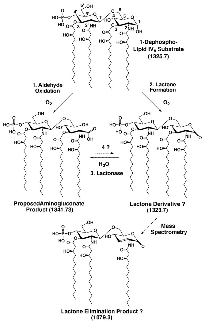
The structures of Rhizobium leguminosarum and Rhizobium etli lipid A are distinct from those found in other Gram-negative bacteria. Whereas the more typical Escherichia coli lipid A is a hexa-acylated disaccharide of glucosamine that is phosphorylated at positions 1 and 4', R. etli and R. leguminosarum lipid A consists of a mixture of structurally related species (designated A-E) that lack phosphate. A conserved distal unit, comprised of a diacylated glucosamine moiety with galacturonic acid residue at position 4' and a secondary 27-hydroxyoctacosanoyl (27-OH-C28) as part of a 2' acyloxyacyl moiety, is present in all five components. The proximal end is heterogeneous, differing in the number and lengths of acyl chains and in the identity of the sugar itself. A proximal glucosamine unit is present in B and C, but an unusual 2-amino-2-deoxy-gluconate moiety is found in D-1 and E. We now demonstrate that membranes of R. leguminosarum and R. etli can convert B to D-1 in a reaction that requires added detergent and is inhibited by EDTA. Membranes of Sinorhizobium meliloti and E. coli lack this activity. Mass spectrometry demonstrates that B is oxidized in vitro to a substance that is 16 atomic mass units larger, consistent with the formation of D-1. The oxidation of the lipid A proximal unit is also demonstrated by matrix-assisted laser desorption ionization time-of-flight mass spectrometry in the positive and negative modes using the model substrate, 1-dephospho-lipid IV(A). With this material, an additional intermediate (or by product) is detected that is tentatively identified as a lactone derivative of 1-dephospho-lipid IV(A). The enzyme, presumed to be an oxidase, is located exclusively in the outer membrane of R. leguminosarum as judged by sucrose gradient analysis. To our knowledge, an oxidase associated with the outer membranes of Gram-negative bacteria has not been reported previously.


Figures

FIG. 1
FIG. 1. Structures of the predominant lipid A species in E. coli, R. etli, and R. leguminosarum
R. etli lipid A, which is thought to be the same as that of R. leguminosarum, is a mixture of several related species (–25). In contrast to E. coli lipid A, all R. etli lipid A species lack phosphate groups (24, 25). Instead, each one contains a galacturonic acid moiety at position 4′ and a single acyloxyacyl unit, featuring an unusual 27-OH-C28 secondary acyl chain, at position 2′ (24, 25). The dashed bonds indicate partial substitutions in the major R. etli components B and D-1 (24, 25). The molecular weight of the largest, fully substituted form of each component is indicated in parentheses. The minor R. etli lipid A species C and E (not shown) are the 3-O-deacylated derivatives of B and D-1, respectively (24, 25). The proximal sugar is a glucosamine unit in B and an aminogluconate moiety in D-1 (24, 25). The proximal 3-deoxy-d-manno-2-octulosonic acid (Kdo) residue of the core oligosaccharide (not present in lipid A prepared by mild acid hydrolysis) is attached at position 6′ in intact LPS.
FIG. 2
FIG. 2. Conversion of [14C]B to [14C]D-1 by R. leguminosarum membranes
The substrate [14C]B, prepared from R. etli cells labeled with [U-14C]acetate (24, 25), was converted to a substance that migrates like [14C]D-1 in the presence of R. leguminosarum 3855 membranes and 0.1% Triton X-100 when analyzed on silica gel TLC plates with the solvent system CHCl3, MeOH, H2O, pyridine (40:25:4:2, v/v). All of the incubations were at 30 °C for 1 h at a protein concentration of 0.5 mg/ml in a reaction mixture containing 50 mm MES, pH 6.5, 0.1% Triton X-100, and 10 µm substrate. Recombining the cytosol and washed membranes did not stimulate the reaction (not shown). S.F., solvent front.
FIG. 3
FIG. 3. Time dependence of the [14C]B to [14C]D-1 conversion
Using 10 µm [14C]B under standard assay conditions, portions of a reaction mixture containing 0.25 mg/ml R. leguminosarum 3855 membranes were spotted on a silica gel TLC plate at various times (0, 15, 30, 60, 90, and 120 min), and the gel was developed in the solvent CHCl3, MeOH, H2O, pyridine (40:25:4:2, v/v) and analyzed with a PhosphorImager, as shown in the upper panel. The quantification of the conversion of B to D-1 is shown in the lower panel. Product formation was linear with protein concentration for 15 min between 0.1 and 0.5 mg/ml membrane protein (data not shown).
FIG. 4
FIG. 4. Inhibition of conversion of [14C]B to [14C]D-1 by EDTA and reactivation with excess Mg2+ and some other divalent cations
A, the conversion of [14C]B to [14C]D-1 (lane 1a) is inhibited by 5 mm EDTA (lane 2a) when added to a reaction mixture containing 50 mm MES, pH 6.5, 0.1% Triton X-100, 10 µm [14C]B, and 0.5 mg/ml R. leguminosarum 3855 membranes. The reaction mixtures in lanes 1a and 2a were incubated for 15 min at 30 °C. B, as shown in lanes 1b and 2b, additional product formation was seen only in the absence of EDTA (lane 1b) when the reaction mixtures from lanes 1a and 2a were incubated for another 20 min at 30 °C. However, when the chloride salts of certain divalent cations were added in excess (10 mm) to a portion of the EDTA inhibited reaction mixture from lane 2a of A and the incubation was allowed to proceed for another 20 min (B), the activity was restored in some cases, as indicated. Product formation was analyzed with a PhosphorImager as for Fig. 2.
FIG. 5
FIG. 5. Conversion of [14C]B to [14C]D-1 by membranes of different R. etli and R. leguminosarum strains
As shown in lanes 1 and 3–5, only R. leguminosarum and R. etli membranes (0.5 mg/ml) can metabolize [14C]B to [14C]D-1. The membranes of E. coli and S. meliloti 1021, which have phosphate-containing lipid A species and do not make 2-aminogluconate, are inactive. The reactions were terminated after 20 min at 30 °C. Product formation was analyzed by TLC, as for Fig. 2 and Fig. 3.
FIG. 6
FIG. 6. MALDI/TOF mass spectrometry of component D-1 synthesized in vitro from B by R. leguminosarum 3855 membranes
R. etli component B (50 µm) was incubated with R. leguminosarum 3855 membranes as described under “Experimental Procedures.” The resulting product that migrated like D-1 during TLC analysis was reisolated and analyzed with a MALDI/TOF mass spectrometer operated in the positive reflectron mode. The partial spectrum of the remaining substrate B is shown in the lower panel. The peaks at m/z 1980.1 and 2008.1 correspond to the expected monoisotopic [M + Na]+ ions for the two major forms of B, which differ by two methylene units (28 atomic mass units), as indicated in Fig. 1 (24). The upper panel shows the same region of the partial spectrum of D-1 synthesized in vitro, which consists of molecules that are all 16 mass units larger than their counterparts in B. The peaks at m/z 1996.5 and 2024.5 correspond to the expected monoisotopic [M + Na]+ ions for the two major forms of D-1, which differ by two methylene groups (28 atomic mass units), consistent with the analysis of the same lipid A molecules isolated from cells, as reported by Que et al. (24).
FIG. 7
FIG. 7. Conversion of [4′-32P]1-dephospho-lipid IVA to several new products by R. leguminosarum 3855 membranes
R. leguminosarum 3855 membranes (0.5 mg/ml) were incubated in parallel for the indicated times with either 10 µm [4′-32P]lipid IV(AA) or [4′-32P]1-dephospho-lipid IVA (B). The [4′-32P]1-dephospho-lipid IVA is rapidly metabolized, whereas the [4′-32P]lipid IVA is not.
FIG. 8
FIG. 8. Hypothetical enzymatic routes for converting 1-dephospho-lipid IVA to a 2-aminogluconate containing species
The expected molecular weights are indicated for each lipid in parentheses. In the first model (reaction 1), the open aldehyde form of the proximal unit of 1-dephospho-lipid IVA would be oxidized to the 2-aminogluconate product. In the second model (reactions 2 and 3), a lactone intermediate is generated by oxidation of the proximal pyranose of 1-dephospho-lipid IVA, followed by lactonase catalyzed hydrolysis to generate the 2-aminogluconate unit. It is conceivable that the oxidation and the lactone opening are both catalyzed by the same protein or that the latter step is nonenzymatic. Although the intact lactone is not detected during mass spectrometry (see below), a fragment with the molecular weight expected for the proposed elimination product of the lactone is in fact very prominent. Furthermore, it also remains a formal possibility that the lactone is formed nonenzymatically from the 2-aminogluconate moiety (reaction 4). In the in vitro conversion of component B to D-1 (Fig. 1 to Fig 6), the putative lactone derivative apparently does not accumulate to the extent seen with 1-dephospho-lipid IVA.
FIG. 9
FIG. 9. Negative-mode MALDI/TOF mass spectrometry of the oxidation products of 1-dephospho-lipid IVA
The lipids extracted from an overnight reaction mixture containing R. leguminosarum 3855 membranes and 1-dephospho-lipid IVA were fractionated on a DEAE-cellulose column. A, the spectrum of the substrate 1-dephospho-lipid IVA prior to incubation with membranes. B, the lipids from the CHCl3, MeOH, 60 mm aqueous NH4Ac (2:3:1, v/v) wash, consisting of residual substrate and the putative lactone. C, the lipids from the CHCl3, MeOH, 120 mm aqueous NH4Ac (2:3:1, v/v) wash. The latter consist primarily of the proposed 2-aminogluconate derivative, as judged by the peak at m/z 1341.2. The peak at m/z 928.9 could not be assigned and may reflect contaminating R. leguminosarum membrane lipids.
FIG. 10
FIG. 10. Positive-mode MALDI/TOF mass spectrometry of the oxidation products of 1-dephospho-lipid IVA
The lipids extracted from an overnight reaction mixture containing R. leguminosarum 3855 membranes and 1-dephospho-lipid IVA were fractionated on a DEAE-cellulose column as in Fig. 9.A, the substrate 1-dephospho-lipid IVA. B, the lipids from the CHCl3, MeOH, 60 mm aqueous NH4Ac (2:3:1, v/v) wash. C, the lipids from the CHCl3, MeOH, 120 mm aqueous NH4Ac (2:3:1, v/v) wash. The peaks at m/z 931.1, 854.0, and 813.4 could not be assigned and may reflect contaminating R. leguminosarum membrane lipids. The B1+ ions of the substrate 1-dephospho-lipid IVA, the lactone elimination product, and the aminogluconate are all observed at m/z 695.3, demonstrating that the distal unit is unchanged and that only the proximal unit of the product is modified under these conditions.
FIG. 11
FIG. 11. Outer membrane localization of the lipid A oxidase in R. leguminosarum 3855
the washed membranes of R. leguminosarum 3855 were separated into inner and outer fractions by isopycnic sucrose density gradient centrifugation (46). Marker enzymes for the outer and inner membranes (phospholipase A2 and NADH oxidase, respectively) were used to confirm the separation. Top panel, turbidity (OD600) was used to detect membrane fragments, and each fraction was also assayed for the R. leguminosarum lipid A oxidase at 30 °C for 30 min under standard conditions by following the conversion of [14C-]B to [14C-]D-1. Lower panel, phospholipase A2 and NADH oxidase marker enzymes.

Similar articles

Cited by

References

    1. Niner BM, Hirsch AM. Symbiosis. 1998;24:51–102.
    1. Stacey G, Burris RH, Evans HJ, editors. Biological Nitrogen Fixation. New York: Routledge, Chapman and Hall, Inc.; 1992.
    1. Triplett EW, editor. Prokaryotic Nitrogen Fixation: A Model System for the Analysis of a Biological Process. Wymondham, UK: Horizon Scientific Press; 2000.
    1. Broughton WJ, Jabbouri S, Perret X. J. Bacteriol. 2000;182:5641–5652. - PMC - PubMed
    1. Downie JA, Walker SA. Curr. Opin. Plant Biol. 1999;2:483–489. - PubMed

Publication types

MeSH terms

LinkOut - more resources