. 2003 Apr 4;278(14):12120-9.
doi: 10.1074/jbc.M300379200. Epub 2003 Jan 15.

Origin of the 2-amino-2-deoxy-gluconate unit in Rhizobium leguminosarum lipid A. Expression cloning of the outer membrane oxidase LpxQ

Affiliations

Origin of the 2-amino-2-deoxy-gluconate unit in Rhizobium leguminosarum lipid A. Expression cloning of the outer membrane oxidase LpxQ

Nanette L S Que-Gewirth et al. J Biol Chem. .

Abstract

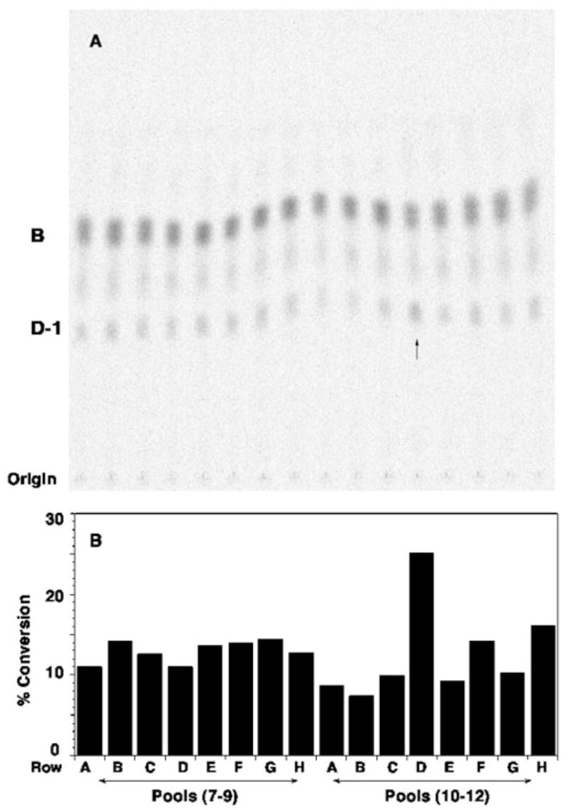
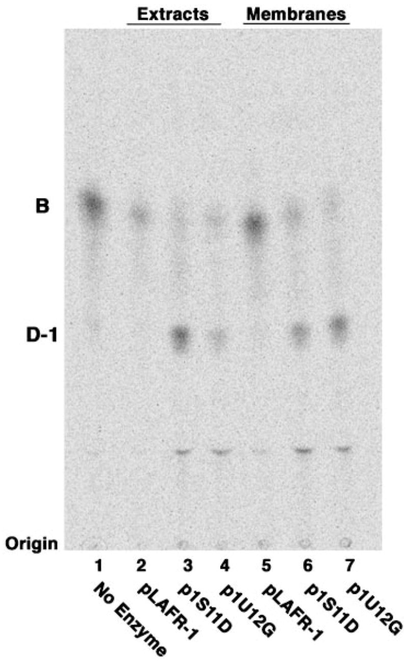
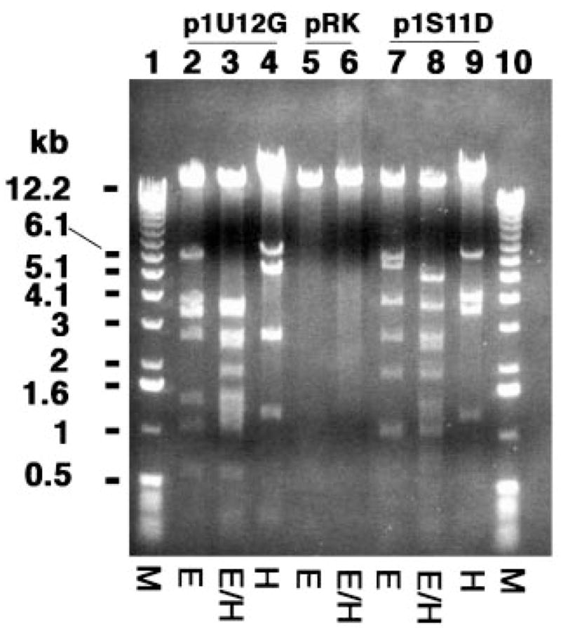
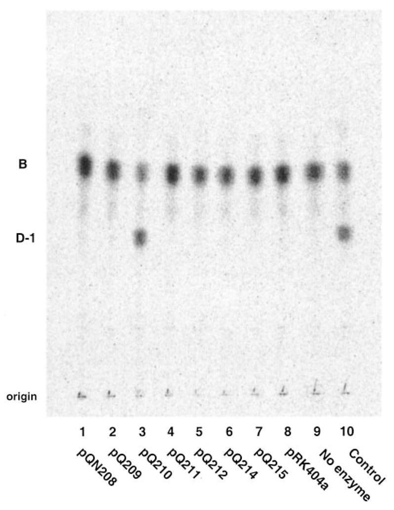
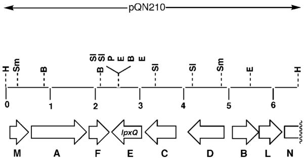
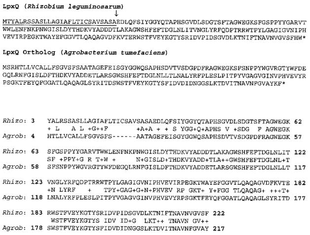
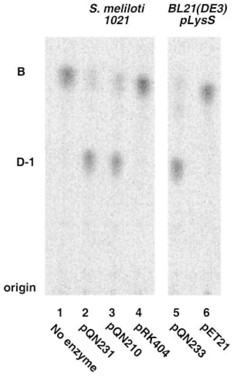
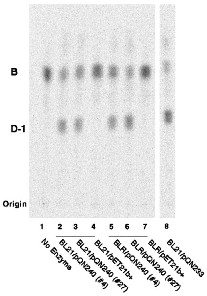
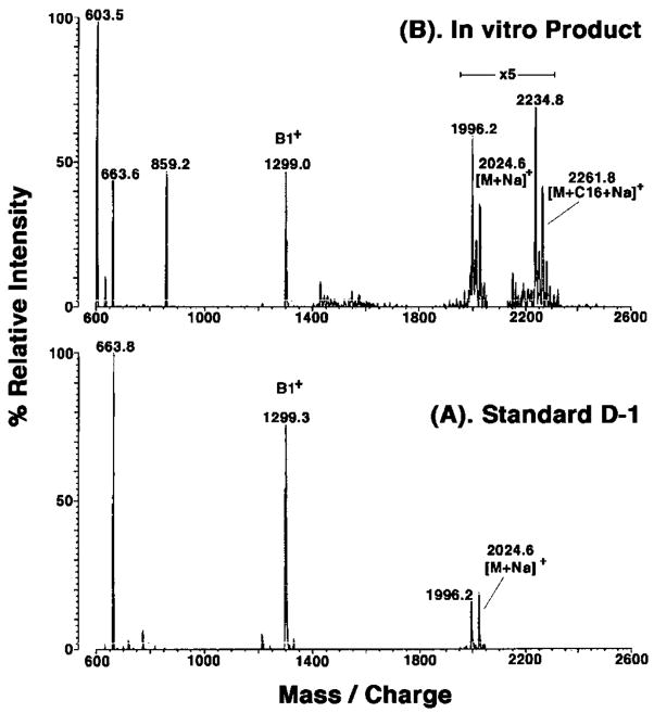
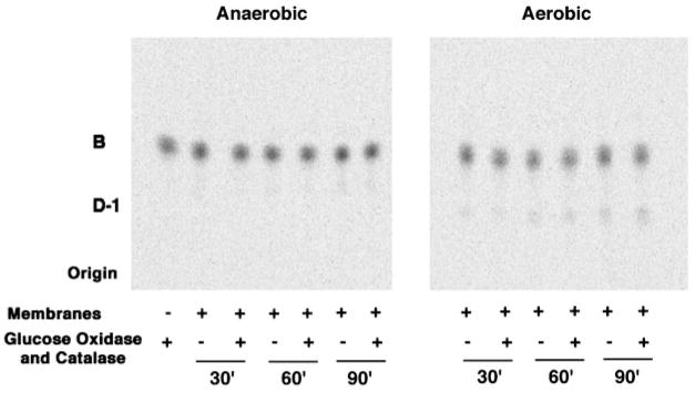
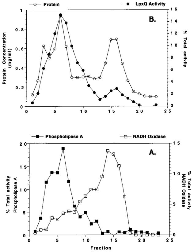
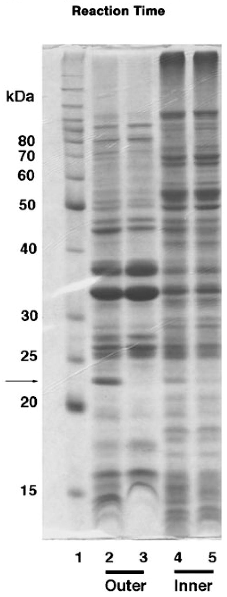
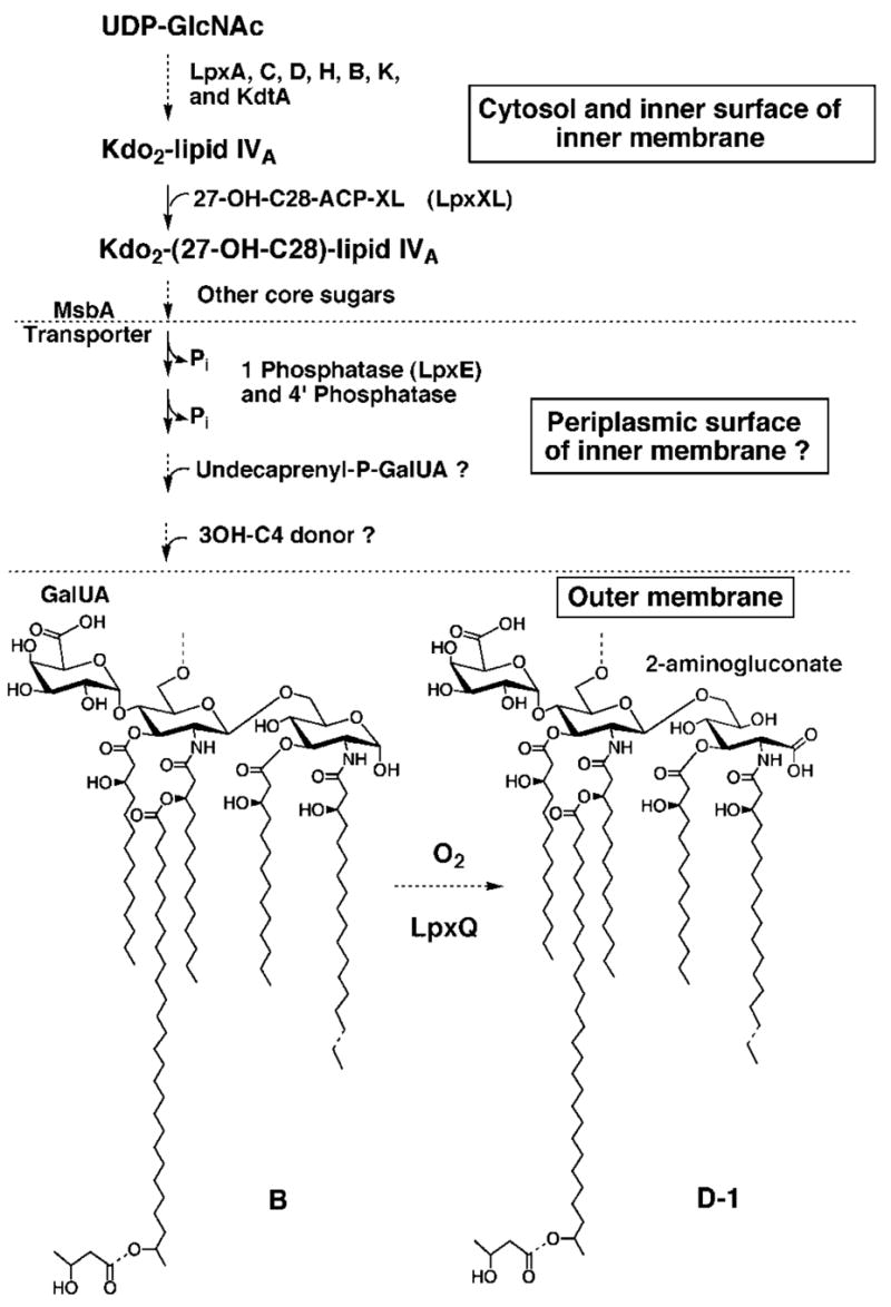
An unusual feature of the lipid A from the plant endosymbionts Rhizobium etli and Rhizobium leguminosarum is the presence of a proximal sugar unit consisting of a 2-amino-2-deoxy-gluconate moiety in place of glucosamine. An outer membrane oxidase that generates the 2-amino-2-deoxy-gluconate unit from a glucosamine-containing precursor is present in membranes of R. leguminosarum and R. etli but not in S. meliloti or Escherichia coli. We now report the identification of a hybrid cosmid that directs the overexpression of this activity by screening 1800 lysates of individual colonies of a R. leguminosarum 3841 genomic DNA library in the host strain R. etli CE3. Two cosmids (p1S11D and p1U12G) were identified in this manner and transferred into S. meliloti, in which they also directed the expression of oxidase activity in the absence of any chromosomal background. Subcloning and sequencing of the oxidase gene on a 6.5-kb fragment derived from the approximately 20-kb insert in p1S11D revealed that the enzyme is encoded by a gene (lpxQ) that specifies a protein of 224 amino acid residues with a putative signal sequence cleavage site at position 28. Heterologous expression of lpxQ using the T7lac promoter system in E. coli resulted in the production of catalytically active oxidase that was localized in the outer membrane. A new outer membrane protein of the size expected for LpxQ was present in this construct and was subjected to microsequencing to confirm its identity and the site of signal peptide cleavage. LpxQ expressed in E. coli generates the same products as seen in R. leguminosarum membranes. LpxQ is dependent on O(2) for activity, as demonstrated by inhibition of the reaction under strictly anaerobic conditions. An ortholog of LpxQ is present in the genome of Agrobacterium tumefaciens, as shown by heterologous expression of oxidase activity in E. coli.


Figures

Fig. 1
Fig. 1. Expression cloning of the R. leguminosarum lipid A oxidase in R. etli
A, a genomic R. leguminosarum DNA library in cosmid pLAFR-1 (24, 32, 44) was transferred into R. etli CE3. Cells from ~1800 cosmid-containing colonies were grown up individually in 96-well microtiter plates. As described under “Experimental Procedures,” the lysates prepared by lysozyme treatment were pooled into groups of three and assayed for overexpression of the lipid A oxidase activity, as judged by the conversion of ~0.003 μM [14C]B (~600 cpm/reaction tube) to [14C]D-1 after 60 min. The arrow indicates a possible pool with elevated oxidase activity. B, the calculated percentage of conversion of [14C]B to [14C]D-1 is shown for each lane. The activity in the pool from row D (wells 10 –12 from plate 1S containing lysates 1S10D, 1S11D, and 1S12D) is approximately twice that of the other pools. Assays of the individual lysates (data not shown) revealed that only 1S11D contained high levels of oxidase activity. Among the ~1800 colonies tested, only three positive cosmids (p1S11D, p1E11D, and p1U12G) were identified in this manner. Based on restriction enzyme digests, p1S11D and p1E11D contained the same insert.
Fig. 2
Fig. 2. Transfer of the R. leguminosarum cosmids expressing the lipid A oxidase activity into S. meliloti
The active cosmids identified in Fig. 1 and the empty vector pLAFR-1 were transferred into S. meliloti 1021 via tri-parental mating (27, 28). Cell-free extracts and membranes derived from late log phase cells containing these cosmids were prepared and assayed for oxidase activity using 10 μM [14C]B (600 cpm/reaction tube) as the substrate. Lanes 2–4 show the results with 0.5 mg/ml crude extracts, whereas lanes 5–7 show assays with 0.5 mg/ml washed membranes. Lane 1, no enzyme control; lane 2, pLAFR-1; lane 3, p1S11D; lane 4, p1U12G; lane 5, pLAFR-1; lane 6, p1S11D; lane 7, p1U12G. The reaction mixtures were incubated overnight at 30 °C. Similar results were obtained when [4′-32P]1-dephospholipid IVA was used as the substrate (data not shown).
Fig. 3
Fig. 3. Digestion of cosmids p1S11D, p1U12G, and pRK404a with EcoRI and HindIII
Cosmids p1U12G (lanes 2–4) and p1S11D (lanes 7–9) were digested with EcoRI (E), HindIII (H), or with both EcoRI and HindIII (E/H). The vector pRK404a was digested in parallel (lanes 5 and 6). The HindIII fragment migrating near 6.5 kb (indicated by the thin line) directed the overexpression of oxidase activity (see below).
Fig. 4
Fig. 4. A 6.5-kb HindIII digestion fragment of p1S11D directs the overexpression of the oxidase
The cosmid p1S11D harbors ~20 kb of R. leguminosarum 8401 genomic DNA. Subclones of the insert were constructed by restriction enzyme digestion and ligation of the fragments into pRK404a. Plasmids pQN209 –pQN214 were derived from various HindIII fragments of the p1S11D insert. pQN208 contains a 3.9-kb EcoRI fragment of the insert, and pQN215 contains a ~0.5-kb PstI fragment. All of the constructs were transferred into S. meliloti 1021 by tri-parental mating (27, 28). Only pQN210, which contains the ~6.5-kb HindIII fragment of the p1S11D insert (see Fig. 3) directed the overexpression of oxidase activity in S. meliloti, as shown by assaying 0.5 mg/ml washed membranes from cells harboring the various constructs with 5 μM [14C]B for 30 min at 30 °C.
Fig. 5
Fig. 5. Order of the R. leguminosarum genes present on the DNA insert in plasmid pQN210
Open reading frames contained within the 6.5-kb insert present in pQN210 were identified based on analysis of the DNA sequence (accession number AY228164) with the program ORF Finder (31) and compared with the nonredundant data base with BLASTx (54). The most plausible candidate for the oxidase is the lpxQ gene. H, HindIII; B, BamHI; Sm, SmaI; Sl, SalI; P, PstI; E, EcoRI.
Fig. 6
Fig. 6. Sequence comparison of LpxQ from R. leguminosarum and Agrobacterium tumefaciens
The predicted amino acid sequence of the R. leguminosarum lipid A oxidase LpxQ (accession number AY228164) is compared with an ortholog of unknown function from A. tumefaciens (17, 18). The order and sequence of the other genes around LpxQ is likewise conserved in both organisms. No other proteins with significant similarity to LpxQ are present in the NCBI data base.
Fig. 7
Fig. 7. Heterologous expression of the lpxQ encoded oxidase in S. meliloti and E. coli
The lpxQ gene was amplified by PCR and ligated into both the shuttle vector pRK404a and into the T7-promoter based vector pET21a+. The resulting constructs, pQN231 and pQN233, were transferred into S. meliloti 1021 and E. coli BL21(DE3)/pLysS, respectively. S. meliloti/pQN231 membranes were prepared from late log phase cells. Membranes of BL21(DE3)/pLysS/pQN233 were obtained from mid log phase cells induced with 1 mM IPTG for 3 h. The membranes (0.5 mg/ml) were assayed for 120 min at 30 °C for their ability to convert 10 μM [14C]B to [14C]D-1 under standard oxidase assay conditions.
Fig. 8
Fig. 8. The lpxQ homolog of A. tumefaciens directs the expression of oxidase activity in E. coli
The lpxQ homolog of A. tumefaciens was amplified by PCR and cloned into pET21b+. The resulting hybrid plasmid is designated pQN240. Membranes of E. coli BL21(DE3)/pLysS/pQN240 (lanes 2 and 3) or BLR(DE3)/pLysS/pQN240 (lanes 5 and 6), grown and induced as described in the legend to Fig. 7, were assayed under standard conditions for 15 min. The no enzyme control is shown in lane 1. Membranes derived from cells containing the vector were assayed in parallel (lanes 4 and 7). A positive control (i.e. membranes of the R. leguminosarum lpxQ overexpressing strain E. coli BL21(DE3)/pLysS/pQN233) is shown in lane 8.
Fig. 9
Fig. 9. Positive ion MALDI/TOF mass spectrum of D-1 generated by membranes of BL21(DE3)/pLysS/pQN233
A reaction mixture (5 ml) containing 50 μM B was incubated for 16 h at 30 °C with 0.5 mg/ml membranes of BL21(DE3)/pLysS/pQN233 under standard conditions, as described in the accompanying article (1). Following partial purification of the D-1-like reaction product by ion exchange chromatography on a small column of DEAE-cellulose, MALDI/TOF mass spectrometry was performed in the positive ion mode (1). Panel A shows the spectrum of a standard preparation of component D-1 isolated from R. etli (3). Panel B shows the spectrum of the partially purified in vitro reaction product. In addition to confirming the incorporation of an oxygen atom into the proximal unit of B, this spectrum also reveals that about half of the D-1 was further modified with a palmitate residue, presumably because of the PagP acyltransferase activity that is present in membranes of the E. coli host strain (7).
Fig. 10
Fig. 10. The lipid A oxidase encoded by lpxQ is dependent upon atmospheric oxygen
The oxidase assay was performed either in an anaerobic chamber or under ambient atmospheric conditions, using membranes of an E. coli strain expressing a C-terminal His-tagged version of LpxQ. The conversion of [14C]B to [14C]D-1 was determined after 30, 60, or 90 min at 30 °C, using 0.1 mg/ml membranes from strain BLR(DE3)/pLysS/pQN235 that was induced with IPTG as in Fig. 5. The presence of glucose oxidase and catalase had little or no effect on the rate of the reaction or the extent of conversion.
Fig. 11
Fig. 11. Outer membrane localization of LpxQ oxidase activity in E. coli Novablue(DE3)/pQN233
Washed membranes obtained from induced cells of E. coli Novablue(DE3)/pQN233 were separated by isopycnic sucrose density gradient centrifugation. Marker enzymes localized in the outer or inner membranes (phospholipase A and NADH oxidase respectively) were assayed to evaluate the extent of separation. A, phospholipase A and NADH oxidase activity. B, LpxQ oxidase activity and protein concentration.
Fig. 12
Fig. 12. Outer membrane localization of overexpressed LpxQ protein in E. coli Novablue(DE3)/pQN233
Membranes of E. coli Novablue(DE3)/pQN233 and Novablue(DE3)/pET21a+ were fractionated by isopycnic sucrose gradient centrifugation as described under “Experimental Procedures.” Peak fractions of outer and inner membranes were pooled separately, and recovered by ultracentrifugation. The membranes were subjected to SDS-PAGE (12% gel and 40 μg of protein/lane). A band of the size expected for LpxQ is seen only in the outer membranes derived from E. coli Novablue(DE3)/pQN233, as indicated by the arrow. Lane 1, Molecular weight marker (Benchmark Protein Ladder from Invitrogen); lane 2, outer membranes of Novablue(DE3)/pQN233; lane 3, outer membranes of Novablue(DE3)/pET21a+; lane 4, inner membranes of Novablue(DE3)/pQN233; lane 5, inner membranes of Novablue(DE3)/pET21a+. The putative LpxQ band, indicated by the arrow, was excised and subjected to N-terminal microsequencing.
Fig. 13
Fig. 13. Proposed compartmentalization of R. leguminosarum lipid A biosynthesis and function of LpxQ
Almost all of the enzymes needed to generate component B have been detected in extracts of R. leguminosarum and R. etli (, , –57). The only exceptions are the reactions that incorporate the 4 ′-galacturonic acid and the β-hydroxybutyrate residues. Although the hypothetical glycolipid, undecaprenylphosphate-GalUA, has not actually been isolated from cells or confirmed as the GalUA donor substrate in vitro, recent work in our laboratory suggests that GalUA transfer to R. leguminosarum lipopolysaccharide precursors requires a membrane-bound donor (S. S. Basu, M. Kanipes, and C. R. H. Raetz, unpublished results). The lpxE gene, which was recently found by expression cloning,2 encodes the 1-phosphatase, and it is predicted to have a periplasmic active site. The gene encoding the 4 ′-phosphatase is unknown. The ABC transporter MsbA is proposed to catalyze the flip-flop of the nascent lipid A 1,4 ′ bis-phosphate with attached core sugars across the inner membrane (–60), thereby presenting this intermediate to the periplasmic lipid A modification enzymes. Following the formation of component B and transport to the outer membrane by unknown mechanisms, the LpxQ oxidase converts the proximal glucosamine residue to the 2-aminogluconate unit in an oxygen-dependent manner. If the oxidation reaction proceeds through a lactone intermediate (as discussed in the accompanying article (1)), an additional lactonase (not shown) might be needed to generate D-1. However, hydrolysis of such a lactone could be catalyzed by LpxQ itself or be nonenzymatic.

Similar articles

Cited by

References

    1. Que-Gewirth NLS, Lin S, Cotter RJ, Raetz CRH. J Biol Chem. 2003;278:12109–12119. - PMC - PubMed
    1. Bhat UR, Forsberg LS, Carlson RW. J Biol Chem. 1994;269:14402–14410. - PubMed
    1. Que NLS, Lin S, Cotter RJ, Raetz CRH. J Biol Chem. 2000;275:28006–28016. - PMC - PubMed
    1. Que NLS, Ribeiro AA, Raetz CRH. J Biol Chem. 2000;275:28017–28027. - PMC - PubMed
    1. Nishijima M, Nakaike S, Tamori Y, Nojima S. Eur J Biochem. 1977;73:115–124. - PubMed

Publication types

MeSH terms

LinkOut - more resources