

Increased diffusion in the brain of professional boxers: a preclinical sign of traumatic brain injury?
- PMID: 12533327
- PMCID: PMC8148951
Increased diffusion in the brain of professional boxers: a preclinical sign of traumatic brain injury?
Abstract
Background and purpose: Professional boxing is associated with chronic, repetitive head blows that may cause brain injuries. Diffusion-weighted imaging is sensitive to microscopic changes and may be a useful tool to quantify the microstructural integrity of the brain. In this study, we sought to quantify microscopic alterations associated with chronic traumatic brain injury in professional boxers.
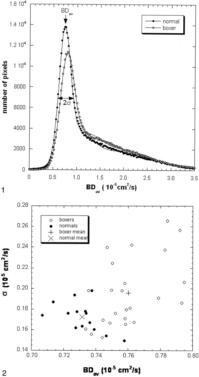
Methods: MR and diffusion-weighted imaging were performed in 24 boxers and in 14 age- and sex-matched control subjects with no history of head trauma. Using distribution analysis, the average diffusion constant of the entire brain (BD(av)) and diffusion distribution width (sigma) were calculated for each subject; findings in professional boxers were compared with those of control subjects. In the boxer group, correlations between diffusion changes and boxing history and diffusion changes and MR imaging findings were assessed.
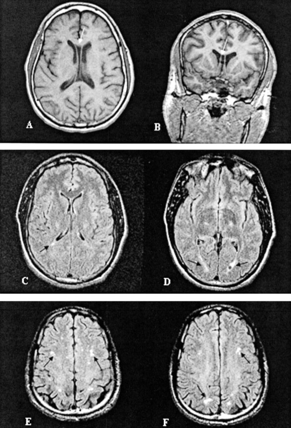
Results: The measured diffusion values in the boxer group were significantly higher than those measured in the control group (BD(av), P <.0001; sigma, P <.01). In the boxer group, a robust correlation was found between increased BD(av) and frequency of hospitalization for boxing injuries (r = 0.654, P <.05). The most common MR finding in the boxer group was volume loss inappropriate to age followed by cavum septum pellucidum, subcortical white matter disease, and periventricular white matter disease.
Conclusion: Boxers had higher diffusion constants than those in control subjects. Our data suggest that microstructural damage of the brain associated with chronic traumatic brain injury may elevate whole-brain diffusion. This global elevation can exist even when routine MR findings are normal.
Figures
References
-
- Meythaler JM, Peduzzi JD, Eleftheriou E, Novack TA. Current concepts: diffuse axonal injury–associated traumatic brain injury. Arch Phys Med Rehabil 2001;82:1461–1471 - PubMed
-
- Moseley IF. The Neuroimaging evidence for chronic brain due to boxing. Neuroradiology 2000;42:1–8 - PubMed
-
- Jordan BD, Jahre C, Hauser WH, et al. CT of 338 active professional boxers. Radiology 1992;2:181–185 - PubMed
-
- Jordan BD, Relkin NR, Ravdin LD, Jacobs AR, Bennett A, Gandy S. Apolipoprotein E epsilon4 associated with chronic traumatic brain injury in boxing. JAMA 1997;278:136–140 - PubMed
MeSH terms
LinkOut - more resources
Full Text Sources
Medical