Contrast-enhanced MR imaging of cerebral fat embolism: case report and review of the literature
- PMID: 12533333
- PMCID: PMC8148954
Contrast-enhanced MR imaging of cerebral fat embolism: case report and review of the literature
Abstract
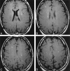
We report a case of cerebral fat embolism (CFE) that was imaged with T2- and T2*-weighted, fluid-attenuated inversion recovery, and contrast-enhanced T1-weighted images. Multiple white matter lesions demonstrated contrast enhancement, with little evidence of hemorrhage. The patient regained complete neurologic function. CFE can cause capillary inflammation and breakdown of the blood-brain barrier, without associated hemorrhage or permanent neurologic deficit. In this case, both contrast-enhanced T1-weighted and gradient-echo T2*-weighted images were important in diagnosing CFE and in determining the severity of the patient's condition.
Figures


References
-
- Johnson MJ, Lucas GL. Fat embolism syndrome. Orthopedics 1996;19:41–47 - PubMed
-
- Gurd AR. Fat embolism: an aid to diagnosis. J Bone Joint Surg 1970;52B:732–737 - PubMed
-
- Takahashi M, Suzuki R, Osakabe Y, et al. Magnetic resonance imaging findings in cerebral fat embolism: correlation with clinical manifestations. J Trauma Inj Infect Crit Care 1999;46:324–327 - PubMed
-
- Kamenar E, Burger PC. Cerebral fat embolism: a neuropathological study of a microembolic state. Stroke 1980;11:477–484 - PubMed
Publication types
MeSH terms
Substances
LinkOut - more resources
Full Text Sources
Medical
