. 2003 Feb;185(3):779-87.
doi: 10.1128/JB.185.3.779-787.2003.

Sizing the holin lesion with an endolysin-beta-galactosidase fusion

Affiliations

Sizing the holin lesion with an endolysin-beta-galactosidase fusion

Ing-Nang Wang et al. J Bacteriol. 2003 Feb.

Abstract

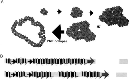
Double-stranded DNA phages require two proteins for efficient host lysis: the endolysin, a muralytic enzyme, and the holin, a small membrane protein. In an event that defines the end of the vegetative cycle, the lambda holin S acts suddenly to permeabilize the membrane. This permeabilization enables the R endolysin to attack the cell wall, after which cell lysis occurs within seconds. A C-terminal fusion of the R endolysin with full-length beta-galactosidase (beta-Gal) was tested for lytic competence in the context of the late-gene expression system of an induced lambda lysogen. Under these conditions, the hybrid R-beta-Gal product, an active tetrameric beta-Gal greater than 480 kDa in mass, was fully functional in lysis mediated by the S holin. Western blot analysis demonstrated that the lytic competence was not due to the proteolytic release of the endolysin domain of the R-beta-Gal fusion protein. The ability of this massive complex to be released by the S holin suggests that S causes a generalized membrane disruption rather than a regular oligomeric membrane pore. Similar results were obtained with an early lysis variant of the S holin and also in parallel experiments with the T4 holin, T, in an identical lambda context. However, premature holin lesions triggered by depolarization of the membrane were nonpermissive for the hybrid endolysin, indicating that these premature lesions constituted less-profound damage to the membrane. Finally, a truncated T holin functional in lysis with the endolysin is completely incompetent for lysis with the hybrid endolysin. A model for the formation of the membrane lesion within homo-oligomeric rafts of holin proteins is discussed.


Figures

FIG. 1.
FIG. 1.
Modification of R with c-myc and lacZ at the 5′ and 3′ termini. Shown are the predicted alterations at the termini of the S and R proteins deriving from the insertions described in the text, in the context of the promoter-proximal region of the λ late transcriptional unit SRRzRz1. The S allele is S105, where the start codon for the S107 antiholin has been eliminated (7). The S′ allele created by the insertion of the c-myc sequence at the start of R has 3 C-terminal residues altered, with a predicted net change of −2 in charge. The c-myc (10 residues) and β-Gal (1,021 residues) sequences are shown in underlined boldface as insertions after codon 1 and before codon 158 of R, respectively.
FIG. 2.
FIG. 2.
The endolysin-β-Gal fusion is fully functional in holin-mediated lysis. Culture growth and lysis were monitored by determining the A550 after thermal inductions. (A) Induction of λΔ(SR) lysogens carrying pS105mycR (circles) or pS105mycRφlacZ (squares). Open symbols, repeat experiment with approximately 1% CHCl3 added at the indicated time; filled symbols, untreated cultures. (B) Induction of MC4100 lysogens carrying λmycR or λmycRφlacZ prophages.
FIG. 3.
FIG. 3.
Plaque-forming ability of λ with the mycRφlacZ hybrid endolysin gene. λmycR and λmycRφlacZ were mixed at a 1:1 ratio and plated with MC4100 in top agar containing X-Gal.
FIG. 4.
FIG. 4.
The lytic function of the hybrid mycR-β-Gal hybrid is not due to proteolytic release of the N-terminal endolysin domain. (A) Full-length mycR and mycR-β-Gal proteins accumulate to equivalent levels. Lysates from the induction of λΔ(SR) lysogens carrying pS105mycR (lane 1) or pS105mycRφlacZ (lane 2) were analyzed by immunoblotting with anti-c-myc antibodies. Values at the left are molecular mass standards (in kilodaltons). (B) The level of mycR endolysin is limiting for the rate of lysis. Culture growth and lysis were monitored by determining the A550 after the induction of λΔ(SR) lysogens carrying pS105mycR (filled circles) or pS105mycRφlacZ (filled squares). Isogenic lysogens carrying pS105R and the compatible plasmid pZS*32-mycR were subjected to lysogenic induction and the addition of either no IPTG (open circles) or 1 mM IPTG (open triangles) at time zero. (C) The levels of R-length proteolytic fragments of the mycR-β-Gal protein were negligible compared to lysis-limiting levels of MycR. Lysates from the induced lysogens carrying pS105mycRφlacZ or pS105R and pZS*32-mycR (no IPTG) were analyzed as described for panel A, except that the stain was deliberately overdeveloped. Values at the right are molecular mass standards (in kilodaltons). (D) Gel filtration chromatography of the mycR and mycR-β-Gal proteins. A lysate prepared from induction of λΔ(SR)pS105mycRφlacZ was analyzed by gel filtration, followed by SDS-PAGE and immunoblotting with anti-c-myc antibodies. Shown is the elution pattern for mass standards as indicated, with the blot being superimposed under the appropriate samples. MAU, mass arbitrary units.
FIG. 5.
FIG. 5.
Abnormal holin lesions can discriminate between the endolysin and the endolysin-β-Gal hybrid. (A) The early-lysis allele of λ S supports lysis with both R and RφlacZ. Culture growth and lysis were monitored by determining the A550 after induction of λΔ(SR) lysogens carrying pS105R (circles) or pS105A52GR (squares). The lysogens also carried a compatible plasmid carrying pZA32-mycR (open symbols) or pZA32-mycRφlacZ (filled symbols). IPTG (1 mM) was added at the time of lysogenic induction (time zero). (B and C) S holin lesions prematurely triggered by energy poisons are differentially permissive for the mycR and mycR-β-Gal endolysins. Culture growth and lysis were monitored by determining the A550 after induction of the λS105mycR (B) or λS105mycRφlacZ (C) lysogen. KCN (10 mM) was added at 20 min (open circles), 30 min (filled circles) or 40 min (open squares). Filled squares, untreated culture. (D) The parental T4 t, but not a t allele with its 69 C-terminal residues deleted, supports lysis with both cmycR and cmycRφlacZ. Conditions were the same as described for panel A except that the plasmid was pT4-t (squares) or pT4-tΔ69 (circles).
FIG. 6.
FIG. 6.
Model for the formation of a holin lesion. (A) Holin rafts. Holins accumulate in rafts in the membrane; intimate helix packing by TMDs largely excludes lipids. Each circle represents a single holin molecule. Spontaneous formation of an aqueous channel by thermal fluctuation is depicted. The localized depolarization causes a conformational change in the holins, leading to asymmetric disruption of the helix packing, exposure of a relatively hydrophilic surface, and dispersion of the subunits into the holin lesion. PMF, proton motive force. (B) Hole size. A schematic representation of the formation of large lesions by the wild-type holin and smaller lesions by a mutant that accretes into more numerous, but smaller, rafts.

References

    1. Bhakdi, S., J. Tranum-Jensen, and A. Sziegoleit. 1985. Mechanism of membrane damage by streptolysin-O. Infect. Immun. 47:52-60. - PMC - PubMed
    1. Bläsi, U., P. Fraisl, C.-Y. Chang, N. Zhang, and R. Young. 1999. The C-terminal sequence of the ë holin constitutes a cytoplasmic regulatory domain. J. Bacteriol. 181:2922-2929. - PMC - PubMed
    1. Bläsi, U., K. Nam, D. Hartz, L. Gold, and R. Young. 1989. Dual translational initiation sites control function of the lambda S gene. EMBO J. 8:3501-3510. - PMC - PubMed
    1. Chang, C.-Y., K. Nam, and R. Young. 1995. S gene expression and the timing of lysis by bacteriophage λ. J. Bacteriol. 177:3283-3294. - PMC - PubMed
    1. Cheng, X., X. Zhang, J. W. Pflugrath, and F. W. Studier. 1994. The structure of bacteriophage T7 lysozyme, a zinc amidase and an inhibitor of T7 RNA polymerase. Proc. Natl. Acad. Sci. USA 91:4034-4038. - PMC - PubMed

Publication types

LinkOut - more resources