. 2003 Feb;185(3):991-1000.
doi: 10.1128/JB.185.3.991-1000.2003.

Characterization of SrgA, a Salmonella enterica serovar Typhimurium virulence plasmid-encoded paralogue of the disulfide oxidoreductase DsbA, essential for biogenesis of plasmid-encoded fimbriae

Affiliations

Characterization of SrgA, a Salmonella enterica serovar Typhimurium virulence plasmid-encoded paralogue of the disulfide oxidoreductase DsbA, essential for biogenesis of plasmid-encoded fimbriae

C W Bouwman et al. J Bacteriol. 2003 Feb.

Abstract

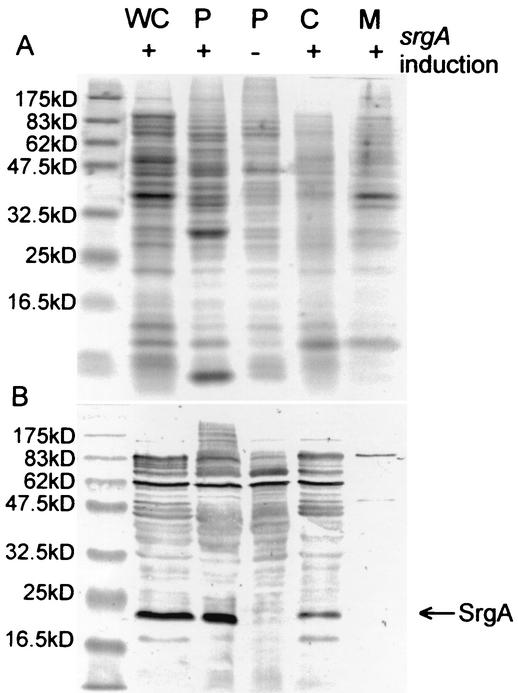
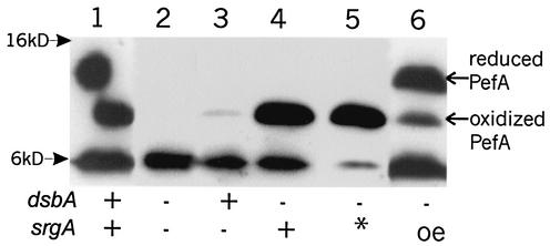
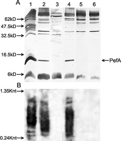
Disulfide oxidoreductases are viewed as foldases that help to maintain proteins on productive folding pathways by enhancing the rate of protein folding through the catalytic incorporation of disulfide bonds. SrgA, encoded on the virulence plasmid pStSR100 of Salmonella enterica serovar Typhimurium and located downstream of the plasmid-borne fimbrial operon, is a disulfide oxidoreductase. Sequence analysis indicates that SrgA is similar to DsbA from, for example, Escherichia coli, but not as highly conserved as most of the chromosomally encoded disulfide oxidoreductases from members of the family Enterobacteriaceae. SrgA is localized to the periplasm, and its disulfide oxidoreductase activity is dependent upon the presence of functional DsbB, the protein that is also responsible for reoxidation of the major disulfide oxidoreductase, DsbA. A quantitative analysis of the disulfide oxidoreductase activity of SrgA showed that SrgA was less efficient than DsbA at introducing disulfide bonds into the substrate alkaline phosphatase, suggesting that SrgA is more substrate specific than DsbA. It was also demonstrated that the disulfide oxidoreductase activity of SrgA is necessary for the production of plasmid-encoded fimbriae. The major structural subunit of the plasmid-encoded fimbriae, PefA, contains a disulfide bond that must be oxidized in order for PefA stability to be maintained and for plasmid-encoded fimbriae to be assembled. SrgA efficiently oxidizes the disulfide bond of PefA, while the S. enterica serovar Typhimurium chromosomally encoded disulfide oxidoreductase DsbA does not. pefA and srgA were also specifically expressed at pH 5.1 but not at pH 7.0, suggesting that the regulatory mechanisms involved in pef gene expression are also involved in srgA expression. SrgA therefore appears to be a substrate-specific disulfide oxidoreductase, thus explaining the requirement for an additional catalyst of disulfide bond formation in addition to DsbA of S. enterica serovar Typhimurium.


Figures

FIG. 1.
FIG. 1.
Comparison of amino acid sequences of SrgA. The stars indicate identical residues between SrgA and DsbA (row &) or between all of the sequences (row all). The A and B domain labeling is based on the crystal structure determination for E. coli DsbA (48), and the B domain is inserted into the A domain in this linear representation. The α-helical (dashed) and β-sheet (dotted) regions are boxed according to Guddat et al. (29). The α-helix region containing the active site is represented in bold. Por is the disulfide oxidoreductase from Haemophilus influenzae (69), and TcpG is the disulfide oxidoreductase from Vibrio cholerae (54).
FIG. 2.
FIG. 2.
SDS-PAGE Coomassie-stained gel (A) and Western immunoblot (B) showing the location of SrgA. Strain NLM2198/pLMN107 was grown in the absence (−) and presence (+) of arabinose induction of SrgA. Cells were harvested and fractionated as described in Materials and Methods. WC, whole-cell lysate; P, periplasmic fraction; C, soluble cytoplasmic fraction; M, membrane fraction. Molecular size markers are indicated to the left of the panels.
FIG. 3.
FIG. 3.
Electron micrographs of negatively stained E. coli MC1061 expressing DsbA (dsbA+) (left column) or not expressing DsbA (dsbA::Km) (right column) with different plasmid constructs in each row as indicated. Panels A and B, pef+ srgA+; panels C and D, pef+ srgA; panels E and F, pef srgA dsbA overexpressed; panels G and H, vector-only control.
FIG. 4.
FIG. 4.
Western immunoblot analysis of SrgA dependence of oxidation status of PefA. Blot was developed with anti-PefA antibodies. Half of the PefA protein in lane 1 shifted upwards to run at the reduced location as a result of dithiothreitol diffusion from the markers in the adjacent lane (not shown). Lane 1, NLM2153/p30; lane 2, NLM2198/p30Tn2.7; lane 3, NLM2153/p30Tn2.7; lane 4, NLM2198/p30; lane 5, NLM2198/p30Tn2.7/pCB6; lane 6, NLM2198/p30Tn2.7/pCB6 with arabinose induction. The presence or absence of dsbA and srgA is indicated at the bottom of the figure (* indicates the presence of srgA in trans; oe indicates overexpression of srgA). The anti-PefA antibody cross-reacts with material running at the dye front of the gel (6-kDa marker). The relative positions of the 16- and 6-kDa molecular size markers are indicated.
FIG. 5.
FIG. 5.
Comparison of growth conditions affecting expression of PefA and transcription of srgA. Samples were loaded in the same order for panels A and B. Panel A, Western immunoblot with anti-PefA antibodies. Molecular size standards are indicated to the left of the image. Panel B, Northern blot with srgA probe. RNA standards are indicated in kilonucleotides (Knt) to the left of the image. Lane 1, NLM2153 containing p30, grown in LB at pH 5.1; lane 2, NLM2217 grown in LB at pH 5.1; lane 3, NLM2217 grown in LB at pH 7.0; lane 4, NLM2217 grown in LB plus glucose at pH 5.1; lane 5, NLM2217 grown in LB plus glucose at pH 7.0; lane 6, NLM2153 grown in LB at pH 5.1.

References

    1. Ahmer, B., J. van Reeuwijk, C. Timmers, P. Valentine, and F. Heffron. 1998. Salmonella typhimurium encodes an SdiA homolog, a putative quorum sensor of the LuxR family that regulates genes on the virulence plasmid. J. Bacteriol. 180:1185-1193. - PMC - PubMed
    1. Bardwell, J. C. A. 1994. Building bridges: disulfide bond formation in the cell. Mol. Microbiol. 14:199-205. - PubMed
    1. Bardwell, J. C. A., J. O. Lee, G. Jander, N. Martin, D. Belin, and J. Beckwith. 1993. A pathway for disulfide bond formation in vivo. Proc. Natl. Acad. Sci. USA 90:1038-1042. - PMC - PubMed
    1. Bardwell, J. C. A., K. McGovern, and J. Beckwith. 1991. Identification of a protein required for disulfide bond formation in vivo. Cell 67:581-589. - PubMed
    1. Baumler, A. J., and F. Heffron. 1995. Identification and sequence analysis of lpfABCDE, a putative fimbrial operon of Salmonella typhimurium. J. Bacteriol. 177:2087-2097. - PMC - PubMed

Publication types

MeSH terms

LinkOut - more resources