. 2003 Feb;185(3):1027-36.
doi: 10.1128/JB.185.3.1027-1036.2003.

Rhamnolipid surfactant production affects biofilm architecture in Pseudomonas aeruginosa PAO1

Affiliations

Rhamnolipid surfactant production affects biofilm architecture in Pseudomonas aeruginosa PAO1

Mary E Davey et al. J Bacteriol. 2003 Feb.

Abstract

In response to certain environmental signals, bacteria will differentiate from an independent free-living mode of growth and take up an interdependent surface-attached existence. These surface-attached microbial communities are known as biofilms. In flowing systems where nutrients are available, biofilms can develop into elaborate three-dimensional structures. The development of biofilm architecture, particularly the spatial arrangement of colonies within the matrix and the open areas surrounding the colonies, is thought to be fundamental to the function of these complex communities. Here we report a new role for rhamnolipid surfactants produced by the opportunistic pathogen Pseudomonas aeruginosa in the maintenance of biofilm architecture. Biofilms produced by mutants deficient in rhamnolipid synthesis do not maintain the noncolonized channels surrounding macrocolonies. We provide evidence that surfactants may be able to maintain open channels by affecting cell-cell interactions and the attachment of bacterial cells to surfaces. The induced synthesis of rhamnolipids during the later stages of biofilm development (when cell density is high) implies an active mechanism whereby the bacteria exploit intercellular interaction and communication to actively maintain these channels. We propose that the maintenance of biofilm architecture represents a previously unrecognized step in the development of these microbial communities.
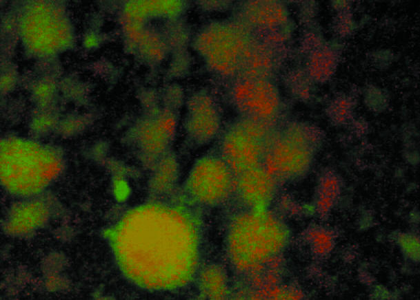

Figures

FIG. 1.
FIG. 1.
A rhamnolipid mutant produces biofilms lacking characteristic architecture. (A) Biofilm formation by the wild type and rhlA mutant in microtiter dishes. The biofilm formation phenotype of the wild type (solid bars) and rhlA mutant (open bars) was quantitated over 24 h by using the microtiter dish assay, as reported elsewhere (26). The A550 value represents crystal violet-stained bacteria attached to the walls of the microtiter dish and is an indirect measure of the biofilm formed. The data represent two experiments, each performed in triplicate. (B) Flow cell architecture of mature biofilms. The wild-type and rhlA::Tn5 strains carrying plasmid pSMC21, which constitutively expresses GFP, were inoculated into flow cells fed by minimal salts EPRI medium supplemented with glucose as the carbon and/or energy source. The top-down views shown here were acquired by epifluorescent microscopy by using the ×63 objective lens, which was used to monitor biofilm development in flow cells over 6 days. The large green macrocolonies are surrounded by dark areas, which are the sparsely colonized channel regions. At day 4, there is no difference between the wild type and the rhlA mutant, thus demonstrating that rhamnolipids are not required for the formation of macrocolonies. For the rhlA mutant, partial filling of the channels can be observed by day 5 and the channels are completely filled in this strain by day 6. The wild-type strain at day 6 displays the characteristic architecture of a mature P. aeruginosa biofilm. Scale bars are included and labeled on the figure. (C) Flow cell architecture of mature biofilms visualized under low magnification. This panel shows a day 6 biofilm obtained at low magnification (with a ×10 objective lens versus the ×63 objective lens used for the images in panel B). Scale bars are included and are labeled on the figure.
FIG. 1.
FIG. 1.
A rhamnolipid mutant produces biofilms lacking characteristic architecture. (A) Biofilm formation by the wild type and rhlA mutant in microtiter dishes. The biofilm formation phenotype of the wild type (solid bars) and rhlA mutant (open bars) was quantitated over 24 h by using the microtiter dish assay, as reported elsewhere (26). The A550 value represents crystal violet-stained bacteria attached to the walls of the microtiter dish and is an indirect measure of the biofilm formed. The data represent two experiments, each performed in triplicate. (B) Flow cell architecture of mature biofilms. The wild-type and rhlA::Tn5 strains carrying plasmid pSMC21, which constitutively expresses GFP, were inoculated into flow cells fed by minimal salts EPRI medium supplemented with glucose as the carbon and/or energy source. The top-down views shown here were acquired by epifluorescent microscopy by using the ×63 objective lens, which was used to monitor biofilm development in flow cells over 6 days. The large green macrocolonies are surrounded by dark areas, which are the sparsely colonized channel regions. At day 4, there is no difference between the wild type and the rhlA mutant, thus demonstrating that rhamnolipids are not required for the formation of macrocolonies. For the rhlA mutant, partial filling of the channels can be observed by day 5 and the channels are completely filled in this strain by day 6. The wild-type strain at day 6 displays the characteristic architecture of a mature P. aeruginosa biofilm. Scale bars are included and labeled on the figure. (C) Flow cell architecture of mature biofilms visualized under low magnification. This panel shows a day 6 biofilm obtained at low magnification (with a ×10 objective lens versus the ×63 objective lens used for the images in panel B). Scale bars are included and are labeled on the figure.
FIG. 2.
FIG. 2.
Partial rescue of the rhlA mutant architecture phenotype by coculture with the wild-type strain. A mixing experiment was done to evaluate the ability of the wild type to rescue the phenotype of the rhlA mutant. The image is a top-down epifluorescent view of a 6-day-old biofilm formed in a flow cell where wild-type cells was mixed 50:50 with the rhlA mutant obtained with a ×10 objective lens. The rhlA mutants cells were visualized by CTC staining (red cells, see Materials and Methods), and the wild-type strain expresses the GFP from a plasmid (green cells). Regions of overlapping wild-type and mutant bacteria are yellow.
FIG. 3.
FIG. 3.
Expression pattern of an rhlA::gfp transcriptional fusion during biofilm formation. The images are top-down views of individual cells and colonies at day 1 (A) and day 2 (B) for flow cell-grown bacteria (obtained with the ×63 objective lens). Both phase-contrast and fluorescent images of the same view are shown. Small clusters of bacteria produced no detectable fluorescence; however, larger colonies (high cell density) resulted in expression of the rhlA::gfp fusion.
FIG. 4.
FIG. 4.
Effect of rhamnolipid overexpression on biofilm formation. The images are fluorescent top-down views of flow cell-grown biofilms formed by wild type and the rhamnolipid mutant at days 1 and 3. PPGAS medium, a low-phosphate medium, induced the synthesis of high levels of rhamnolipids in the wild-type strain and resulted in a sparsely colonized surface. The rhlA mutant was unable to synthesize any rhamnolipids even in low-phosphate medium and made a dense biofilm by day 3.
FIG. 5.
FIG. 5.
Effect of purified rhamnolipids on cell-surface and cell-cell interactions. (Upper panels) Cell-surface interactions. The ability to form a biofilm on polyvinylchloride plastic in the presence or absence of rhamnolipids was examined. The rhamnolipid was added at the time of inoculation, and the biofilm formation was examined after 8 h of incubation. The addition of 250 μM rhamnolipid completely blocked biofilm formation (right panel). (Lower panels) Cell-cell interactions. The effect of rhamnolipids on rafts or pellicles of wild-type P. aeruginosa cells was examined. The disruption of cell-to-cell interactions caused by adding rhamnolipids was captured by phase-contrast microscopy immediately after addition of the compound (right panel). The addition of the same volume of water had no effect on biofilm formation or disruption of the rafts (left panels).
FIG. 6.
FIG. 6.
Biofilm invasion assay. A biofilm of non-GFP-labeled wild-type bacteria was allowed to form for 5 days in a flow cell and then stained with CTC. (a and c) The flow was turned off, ca. 107 GFP-labeled wild-type P. aeruginosa cells were injected into the flow cell, and the flow chamber was incubated without flow for 1 h. (b and d) Flow was resumed for 1 h to remove planktonic bacteria, and the biofilm was examined by fluorescence microscopy. A merged composite image of the fluorescence micrographs is shown. The images in panels a and b were captured by using a ×63 objective lens, and the images in panels c and d were captured by using a ×10 objective lens. Scale bars are included in the images.

Comment in

References

    1. Bloemberg, G. V., G. A. O'Toole, B. J. J. Lugtenberg, and R. Kolter. 1997. Green fluorescent protein as a marker for Pseudomonas spp. Appl. Environ. Microbiol. 63:4543-4551. - PMC - PubMed
    1. Branda, S. S., J. E. Gonzalez-Pastor, S. Ben-Yehuda, R. Losick, and R. Kolter. 2001. Fruiting body formation by Bacillus subtilis. Proc. Natl. Acad. Sci. USA 98:11621-11626. - PMC - PubMed
    1. Brint, J. M., and D. E. Ohman. 1995. Synthesis of multiple exoproducts in Pseudomonas aeruginosa is under the control of RhlR-RhlI, another set of regulators in strain PAO1 with homology to the autoinducer-responsive LuxR-LuxI family. J. Bacteriol. 177:7155-7163. - PMC - PubMed
    1. Christensen, B. B., C. Sternberg, J. B. Andersen, R. J. Palmer, Jr., A. T. Nielsen, M. Givskov, and S. Molin. 1999. Molecular tools for study of biofilm physiology. Methods Enzymol. 310:20-42. - PubMed
    1. Costerton, J. W., P. S. Stewart, and E. P. Greenberg. 1999. Bacterial biofilms: a common cause of persistent infections. Science 284:318-322. - PubMed

Publication types

MeSH terms