. 2003 Jan 15;17(2):240-55.
doi: 10.1101/gad.1032503.

c-MYC apoptotic function is mediated by NRF-1 target genes

Affiliations

c-MYC apoptotic function is mediated by NRF-1 target genes

Fionnuala Morrish et al. Genes Dev. .

Abstract

A detailed understanding of the signaling pathways by which c-Myc elicits apoptosis has proven elusive. In the current study, we have evaluated whether the activation of the mitochondrial apoptotic signaling pathway is linked to c-Myc induction of a subset of genes involved in mitochondrial biogenesis. Cytochrome c and other nuclear-encoded mitochondrial genes are regulated by the transcription factor nuclear respiratory factor-1 (NRF-1). The consensus binding sequence (T/C)GCGCA(C/T)GCGC(A/G) of NRF-1 includes a noncanonical CA(C/T)GCG Myc:MAX binding site. In this study, we establish a link between the induction of NRF-1 target genes and sensitization to apoptosis on serum depletion. We demonstrate, by using Northern analysis, transactivation assays, and in vitro and in vivo promoter binding assays that cytochrome c is a direct target of c-Myc. Like c-Myc, NRF-1 overexpression sensitizes cells to apoptosis on serum depletion. We also demonstrate that selective interference with c-Myc induction of NRF-1 target genes by using a dominant-negative NRF-1 prevented c-Myc-induced apoptosis, without affecting c-Myc-dependent proliferation. These results suggest that c-myc expression leads to mitochondrial dysfunction and apoptosis by deregulating genes involved in mitochondrial function.


Figures
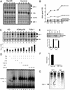
Figure 1
Figure 1
c-Myc transactivation of the NRF-1 target gene cytochrome c. (A) Northern analysis of Myc-ERTM and vector control samples isolated at 0, 2, 4, 6, and 8 h after 4-OHT treatment in the presence of 10 μg/mL cycloheximide. Cells had been serum depleted for 48 h prior to cycloheximide and 4-OHT addition. There are three cytochrome c transcripts of 1.4, 1.1, and 0.7 kb. (B) Graph of the relative level of induction of mRNA over time in Myc-ERTM and vector control lines. Data is corrected for loading by using 18S. This data is representative of three separate experiments. (C) Induction of cytochrome c on serum addition in c-myc−/− and c-myc+/+ cell lines after 48 h of serum depletion. This experiment was repeated twice with similar results. (D) EMSA assay of the ability of Myc:MAX and MAX:MAX to bind to the NRF-1 binding site in the cytochrome c promoter. Myc and MAX were both recombinant forms and used alone at a concentration of 25 ng Myc (lane 2) and 7.5 ng MAX (lane 3) or were combined (lane 4). Samples 5–14 show competition with unlabeled dsDNA samples, including E box (lanes 5,6) a mutated E box (lanes 7,8), wild-type cytochrome c (lanes 911), and mutant cytochrome c DNA (lanes 1214). (E) PCR of input chromatin for c-myc−/− and c-myc+/+ and of chromatin immunoprecipitated with c-Myc antibody, nonimmune serum (mock), and no DNA by using primers for cytochrome c, COX5b, and the negative control albumin. The histograms denote the level of Myc association with cytochrome c and COX5b compared with the albumin control for three replicas. (F) Histograms representing the ability of c-Myc to regulate the cytochrome c promoter through the NRF-1 site or a mutant site in which CATGCG has been mutated to CATTAG. Transfections were done in triplicate, and results are expressed as the fold increase in luciferase activity compared with the empty vector control (pRcCMV) corrected for transfection efficiency, and are representative of four separate experiments. (G) Immunoprecipitation with cytochrome c demonstrates increases in the level of cytochrome c protein synthesis on the activation of c-MYC in serum-deprived cells. Control and MycER cell lysates were immunoprecipitated in the presence of mouse IgG conjugated to agarose or cytochrome c coupled to protein G-Sepharose.
Figure 2
Figure 2
Overexpression of the transcription factor NRF-1 induces apoptosis in serum-depleted cells. (A) Western analysis for three clones expressing the NRF-1-ER fusion protein. (B) Northern analysis of clones overexpressing NRF-1 over a time course of 24 h in the presence of 4-OHT and cycloheximide. (C) Immunoprecipitation with cytochrome c demonstrates increases in the level of cytochrome c protein synthesis in NRF-1-expressing cells under serum deprivation. Control and NRF-1 cell lysates were immunoprecipitated in the presence of mouse IgG conjugated to agarose or cytochrome c coupled to protein G-Sepharose. (D) PI FACS analysis indicating the percentage of sub-G1 cell populations found in three independently derived cell lines overexpressing NRF-1 after 24 h in 0.5% serum. Approximately 105 cells were analyzed in triplicate for each sample, and these results are representative of three separate experiments. (E) Electron microscopy analysis of cells from control and NRF-1-overexpressing cells after 8 h in 0.5% serum. Bars: left and middle panels, 2 μm; right panel, 200 nm.
Figure 3
Figure 3
The antiapoptotic protein Bcl-2 protects cells from NRF1-induced apoptosis. (A) PI FACS analysis of vector control and NRF-1- and Bcl-2/NRF-1-expressing cell lines, indicating the percentage of sub-G1 cells in the population after 24 h in 0.5% serum. A minimum of 105 cells were analyzed in triplicate for each sample, and these results are representative of two separate experiments. (B) Electron microscopy analysis of NRF-1-ERTM and Bcl-2/NRF-1-ERTM cell lines after 24 h in 0.5% serum. Bars, 2 nM.
Figure 4
Figure 4
Down-regulation of NRF-1 target gene transcription on expression of DNRF-1. (A) Northern analysis of NRF-1 target gene expression in Myc-ERTM and DNNRF-1-ERTM/Myc-ERTM cell lines after 4 h of induction with 4-OHT in the presence of 10 μg/mL cycloheximide. (B) Bar graphs illustrating the level of gene suppression in the DNNRF1-ERTM/Myc-ERTM cell line compared with the Myc-ERTM cell line. The white and black bars indicate the level of gene expression at 0 and 4 h, respectively, when corrected for loading by using 18S.
Figure 5
Figure 5
Introduction of DNNRF1 into Myc-ERTM cells inhibits c-Myc-induced apoptosis but maintains cell cycle induction and cell proliferation. (A) PI FACS analysis of vector control, Myc-ERTM, and a DNNRF-1-ERTM/Myc-ERTM cell line, indicating the percentage of sub-G1 cells in the population 48 h after 4-OHT addition. A minimum of 105 cells were analyzed in triplicate for each sample, and these results are representative of three separate experiments. (B) Western analysis using caspase-3 antibody to detect cleavage of caspase 3. (C) PI FACS analysis of the cell cycle in the Myc-ERTM cell line and two representative DNNRF-1-ERTM/Myc-ERTM clones (#1 and #2) in serum-deprived cells in the presence and absence of 4-OHT. Cells were serum depleted in 0.5% serum for 48 h prior to culture in the presence or absence of 4-OHT for 24 h. A minimum of 105 cells were analyzed in triplicate for each sample, and these results are representative of three separate experiments. (D) Viability analysis at 48 and 72 h after 4-OHT addition. (E) Proliferation analysis at 48 and 72 h after 4-OHT addition. Data are expressed as the relative increase in cell proliferation on the addition of 4-OHT compared with cells maintained in the absence of 4-OHT.
Figure 6
Figure 6
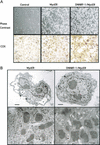
Cellular morphology, ultrastructural, and respiratory changes induced on activation of DNNRF-1 in MycERTM cells. (A) Phase microscopy and COX histochemistry of cells from control, Myc-ERTM, and a DNNRF-1-ERTM/Myc-ERTM clone 48 h after addition of 4-OHT in 0.5% serum. Cells had been serum starved for 48 h prior to 4-OHT addition. (B) Electron micrographs of Myc-ERTM and DNNRF-1-ERTM/MycER-ERTM cells 48 h after 4-OHT addition. The arrows point to mitochondria; asterisks highlight the dilated ER. High-power magnification of mitochondria in Myc-ERTM cells and DNNRF1-ERTM/Myc-ERTM cells illustrates the distinct morphologies of these mitochondria in 0.5% serum. Note that the Myc-ERTM mitochondria have been magnified 2.5× to allow comparison with the mitochondria in the DNNRF1-ERTM/Myc-ERTM cell line. Bars: upper panels, 2 μm; lower panels, 200 nm.
Figure 7
Figure 7
A model for the role of balanced mitochondrial gene expression in the maintenance of cell viability. In the serum-starved cell, quiescence is achieved by creating a balanced expression of genes. On the induction of the transcription factors c-Myc or NRF-1, this balance is disrupted due to the inappropriate expression of a subset of target genes required to maintain mitochondrial function. This leads to mitochondrial dysfunction and death. On the addition of DNNRF-1, a selected subset of genes, which are involved in mitochondrial dysfunction, are down-regulated, allowing the cell to survive. As not all c-Myc target genes are down-regulated, the cell continues to proliferate in the presence of dual signals, which allow survival and proliferation.

Comment in

  • Myc's mastery of mitochondrial mischief.
    Morrish F, Hockenbery D. Morrish F, et al. Cell Cycle. 2003 Jan-Feb;2(1):11-3. doi: 10.4161/cc.2.1.275. Cell Cycle. 2003. PMID: 12695675 Review. No abstract available.

Similar articles

Cited by

References

    1. Bauer M, Schubert A, Rocks O, Grimm S. Adenine nucleotide translocase-1, a component of the permeability transition pore, can dominantly induce apoptosis. J Cell Biol. 1999;147:1493–1501. - PMC - PubMed
    1. Becjker T, Burgess S, Amsterdam A, Allende M, Hopkins N. not really finished is crucial for development of the zebrafish outer retina and encodes a transcription factor highly homologous to human nuclear respiratory factor-1 and avian initiation binding repressor. Development. 1998;125:4369–4378. - PubMed
    1. Bejarano M, Cornvik A, Brijker S, Asker C, Osorio L, Henriksson M. Inhibition of cell growth and apoptosis by inducible expression of the transcriptional repressor Mad1. Exp Cell Res. 2000;260:61–72. - PubMed
    1. Bissonnette R, Echeverri F, Mahboubi A, Green D. Apoptotic cell death induced by c-myc is inhibited by bcl-2. Nature. 1992;359:552–556. - PubMed
    1. Bouchard C, Thieke K, Maier A, Saffrich R, Hanley-Hyde J, Ansorge W, Reed S, Sicinski P, Bartek J, Eilers M. Direct induction of cyclin D2 by Myc contributes to cell cycle progression and sequestration of p27. EMBO J. 1999;18:5321–5333. - PMC - PubMed

Publication types

MeSH terms

LinkOut - more resources