. 2003 Jan 15;17(2):269-81.
doi: 10.1101/gad.1048903.

Tbx1 is regulated by tissue-specific forkhead proteins through a common Sonic hedgehog-responsive enhancer

Affiliations

Tbx1 is regulated by tissue-specific forkhead proteins through a common Sonic hedgehog-responsive enhancer

Hiroyuki Yamagishi et al. Genes Dev. .

Abstract

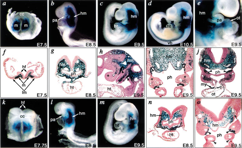
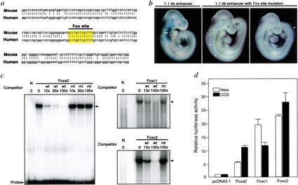
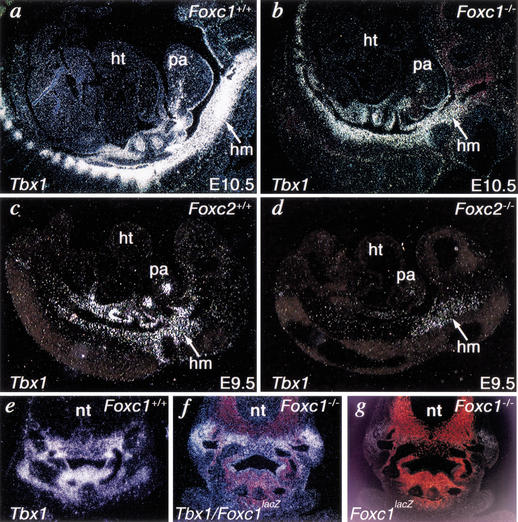
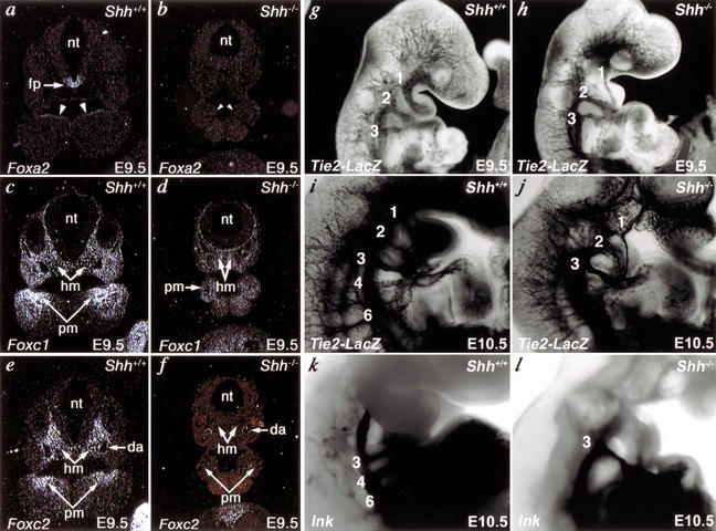
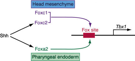
Haploinsufficiency of Tbx1 is likely a major determinant of cardiac and craniofacial birth defects associated with DiGeorge syndrome. Although mice deficient in Tbx1 exhibit pharyngeal and aortic arch defects, the developmental program and mechanisms through which Tbx1 functions are relatively unknown. We identified a single cis-element upstream of Tbx1 that recognized winged helix/forkhead box (Fox)-containing transcription factors and was essential for regulation of Tbx1 transcription in the pharyngeal endoderm and head mesenchyme. The Tbx1 regulatory region was responsive to signaling by Sonic hedgehog (Shh) in vivo. We show that Shh is necessary for aortic arch development, similar to Tbx1, and is also required for expression of Foxa2 and Foxc2 in the pharyngeal endoderm and head mesenchyme, respectively. Foxa2, Foxc1, or Foxc2 could bind and activate transcription through the critical cis-element upstream of Tbx1, and Foxc proteins were required, within their expression domains, for Tbx1 transcription in vivo. We propose that Tbx1 is a direct transcriptional target of Fox proteins and that Fox proteins may serve an intermediary role in Shh regulation of Tbx1.


Figures

Figure 1
Figure 1
Tbx1 expression is controlled by separable pharyngeal endoderm and mesoderm regulatory regions in transgenic mice. (a) Genomic organization of the 5′ mouse Tbx1 locus and flanking region. Boxes indicate exons, and translation start site (ATG) is designated as nucleotide number zero. Construct number is indicated on the left, and the corresponding expression pattern of lacZ at E9.5 is summarized on the right. The far-right column indicates the fraction of F0 transgenic embryos with Tbx1-like lacZ expression/lacZ gene positive embryos. (b,c) Endogenous expression of Tbx1 transcripts is shown by whole-mount RNA in situ hybridization in E9.5 mouse embryos. (dk) Right lateral views of representative embryos obtained with each construct (indicated in upper-right corner of each panel). (c,e,g,i,k) Higher magnification of the pharyngeal arch of embryos in upper panels. Expression of lacZ in embryos with construct 1 (d,e) recapitulated endogenous Tbx1 expression (b,c). LacZ expression in the head mesenchyme (hm) and pharyngeal endoderm (white arrowheads) was detectable in each embryo with construct 1 (e), 3 (g), 4 (i), and 6 (k), whereas that in the pharyngeal mesoderm (black arrowheads) and cardiac outflow tract (ot) was only in embryos with construct 1 (e) or 3 (g). lacZ expression in limb buds (lb) in panel h represents ectopic expression. h, head; ht, heart.
Figure 2
Figure 2
Developmental regulation of Tbx1 expression by tissue-specific enhancers. Embryos from a stable transgenic line harboring the 12.8-kb fragment (construct 1) were analyzed at various times during mouse embryogenesis. (ad) Whole-mount photographs of embryos from E7.5 to E10.5. (e) Right lateral view of embryo focusing on the pharyngeal arch and heart at E9.5. (fj) Transverse (f,g,j), sagittal (h), and frontal (i) sections counterstained by Nuclear Fast Red from E7.5 to E9.5. (a,f) lacZ was expressed in mesoderm cells (m) that give rise to cranial mesenchyme, but not in the cardiac crescent (cc) or lateral plate mesoderm (lm) at E7.5. (b,g) Expression of lacZ was detectable in the head mesenchyme (hm) and pharyngeal arch (pa) at E8.5. (c,h,i,j) lacZ expression was detectable in head mesenchyme, pharyngeal arch mesoderm (white arrowheads), and endoderm (black arrowheads), but not in neural-crest-derived mesenchyme (asterisks) at E9.5. (d) Expression of lacZ extended to the primordia of vertebral bodies (vb). (e,j) lacZ was expressed in both myocardial (my) and endocardial (e) layers of the cardiac outflow tract (ot) at E9.5. (km) Whole-mount photographs of embryos from a stable transgenic line harboring the 1.1-kb fragment (construct 6) at E7.75 to E9.5. (n,o) Transverse (n) or frontal (o) sections of l or m, respectively, counterstained by Nuclear Fast Red. (k) lacZ expression in mesoderm cells was observed similar to panel a at E7.75. (l,n) lacZ was detectable in head mesoderm as in b and g, but excluded from the pharyngeal arch at E8.5. (m,o) Expression of lacZ in head mesenchyme and pharyngeal endoderm was detectable similar to c, i, and j, but expression in pharyngeal mesoderm was absent. hf, head fold; ht, heart; ph, pharynx.
Figure 3
Figure 3
The Tbx1 enhancer is dependent on Shh signaling in a temporo–spatial fashion. (a,b) X-gal staining of wild-type (Shh+/+) and Shh mutant (Shh−/−/) embryos harboring the 12.8-kb Tbx1–lacZ transgene at E9.25 showed relatively normal lacZ expression in the pharyngeal arches and head mesenchyme (hm) of Shh mutant embryos compared with wild-type embryos. (c,d) LacZ expression in wild-type and Shh mutant embryos at E9.5. (e,f) Higher magnification photographs of c and d focusing on pharyngeal arches show down-regulation of lacZ expression in the head mesenchyme and first (I), third (III), and fourth (IV) pharyngeal arches of Shh mutant embryos compared with wild-type embryos. lacZ expression in the second pharyngeal arch (II) was also down-regulated, but expression was still detected in the mesoderm of the second arch of Shh mutant embryos. ht, heart.
Figure 4
Figure 4
A consensus Fox-binding site is essential for regulation of Tbx1. (a) One-hundred percent cross-species conservation of the consensus Fox-binding site at −13,435/−13,423 bp in the 1.1-kb enhancer of Tbx1 is indicated. (b) Mutation of Fox site abolished head mesenchyme (hm) and pharyngeal endoderm (asterisks) enhancer activity in 6/6 lacZ-positive embryos. Ectopic lacZ staining (arrowheads) was observed in some embryos. (c) Electrophoretic gel mobility shift assays (EMSA) revealed DNA–protein complexes of in vitro translated Foxa2, Foxc1, or Foxc2 with the Fox cis-element in the Tbx1 enhancer. The interaction was competed specifically by excess unlabeled wild-type (wt) oligonucleotide, but not by a mutant (mt) oligonucleotide. Fold-excess of competitor DNA compared with labeled probe is shown. No DNA–protein complex (in lane N) was observed with lysate alone. (d) Luciferase expression under control of the Tbx1 genomic DNA fragment shown in panel a after cotransfection of luciferase reporter and Foxa2, Foxc1, or Foxc2 expression construct or empty vector (pcDNA3.1). Results are presented as relative luciferase activity, corrected for transfection efficiency. The control (pcDNA3.1) transfection value is set at 1.
Figure 5
Figure 5
Foxc proteins regulate Tbx1 expression in vivo. (ad) Radioactive in situ hybridization on sagittal sections of E9.5–E10.5 mouse embryos showed that Tbx1 was expressed, but partially down-regulated in the head mesenchyme (hm) of Foxc1 or Foxc2 mutant (Foxc1−/− or Foxc2−/−) embryos compared with wild-type (Foxc1+/+ or Foxc2+/+) embryos. (eg) Transverse section of wild-type or Foxc1 mutants at the pharyngeal arch level. Tbx1 expression (white) in Foxc1-null embryo was down-regulated (f) compared with wild type (e) in the subdomain corresponding to Foxc1 expression, as marked by lacZ expression under control of the endogenous Foxc1 promoter (red in g). Tbx1 expression and lacZ expression are overlaid in f. ht, heart; pa, pharyngeal arch; nt, neural tube.
Figure 6
Figure 6
Shh regulates Fox proteins and aortic arch development. (a,b) Radioactive in situ hybridization on transverse sections of E9.5 mouse embryos shows that Foxa2 is down-regulated in floor plate (fp) of neural tube (nt) and pharyngeal endoderm (arrowheads) of Shh−/− embryos compared with wild-type (Shh+/+) embryos. (c,d) Foxc1 was expressed in the head mesenchyme (hm) and pharyngeal arch mesenchyme (pm) of Shh mutant embryos at relatively low levels compared with wild-type embryos. (e,f) Foxc2 was undetectable in the head mesenchyme and pharyngeal arch mesenchyme of Shh mutant embryos compared with wild-type embryos. Foxc2 expression in dorsal aortae (da) was normal in Shh mutant embryos. a, c, and e are serial sections from a wild-type embryo, and b, d, and f are serial sections from an Shh mutant embryo. (gl), Aortic arch defects in Shh mutants. lacZ driven by the Tie2 promoter marked the vasculature and revealed a small first aortic arch artery at E9.5 (h) and absence of the fourth and sixth arch arteries at E10.5 (j) compared with wild type (g,i). India ink injection demonstrated similar absence of the fourth and sixth arch arteries. Arch arteries are indicated numerically. Right lateral views are shown with close-ups of the pharyngeal arch region in il.
Figure 7
Figure 7
A proposed model for molecular regulation of Tbx1. Our data support a model in which Tbx1 is directly regulated by Foxc1 and Foxc2 in the head mesenchyme, and Foxa2 in the pharyngeal endoderm, through a common Fox-binding site upstream of Tbx1. Shh signaling functions to maintain the expression of Foxa2 and Foxc2, which subsequently activate Tbx1 expression.

References

    1. Abu-Issa R, Smyth G, Smoak I, Yamamura KI, Meyers EN. Fgf8 is required for pharyngeal arch and cardiovascular development in the mouse. Development. 2002;129:4613–4625. - PubMed
    1. Ang SL, Rossant J. HNF-3 β is essential for node and notochord formation in mouse development. Cell. 1994;78:561–574. - PubMed
    1. Bollag RJ, Siegfried Z, Cebra-Thomas JA, Garvey N, Davison EM, Silver LM. An ancient family of embryonically expressed mouse genes sharing a conserved protein motif with the T locus. Nat Genet. 1994;7:383–389. - PubMed
    1. Chapman DL, Garvey N, Hancock S, Alexiou M, Agulnik SI, Gibson-Brown JJ, Cebra-Thomas J, Bollag RJ, Silver LM, Papaioannou VE. Expression of the T-box family genes, Tbx1–Tbx5, during early mouse development. Dev Dyn. 1996;206:379–390. - PubMed
    1. Chiang C, Litingtung Y, Lee E, Young KE, Corden JL, Westphal H, Beachy PA. Cyclopia and defective axial patterning in mice lacking Sonic hedgehog gene function. Nature. 1996;383:407–413. - PubMed

Publication types

MeSH terms