

BETA2/NeuroD1 null mice: a new model for transcription factor-dependent photoreceptor degeneration
- PMID: 12533605
- PMCID: PMC6741880
- DOI: 10.1523/JNEUROSCI.23-02-00453.2003
BETA2/NeuroD1 null mice: a new model for transcription factor-dependent photoreceptor degeneration
Abstract
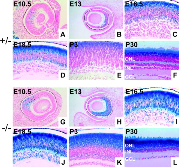
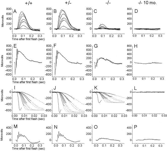
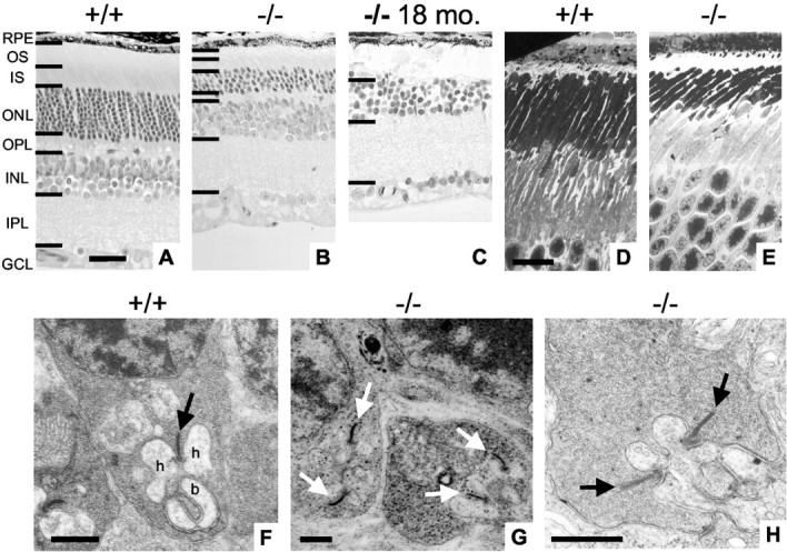
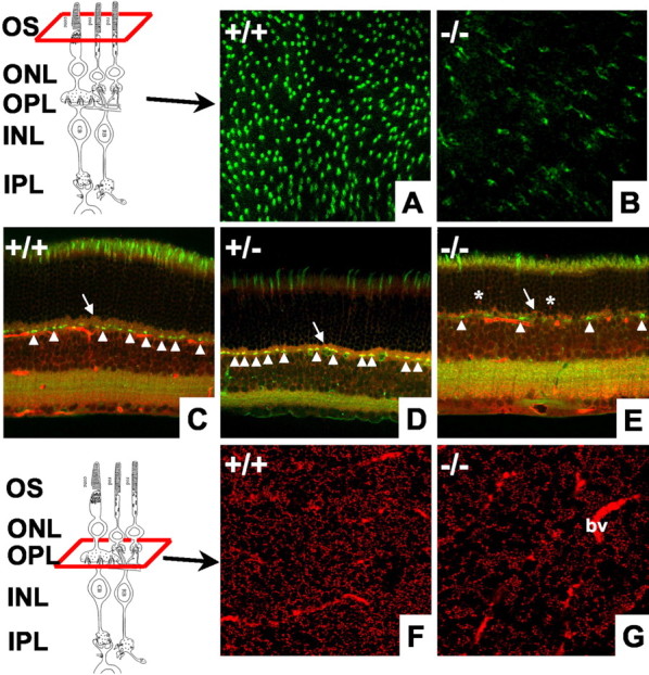
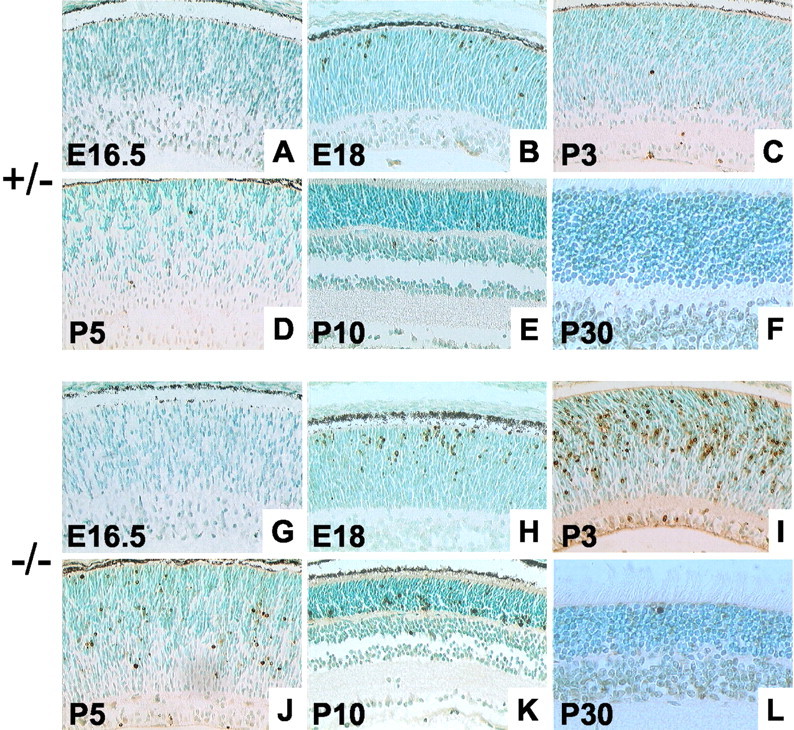
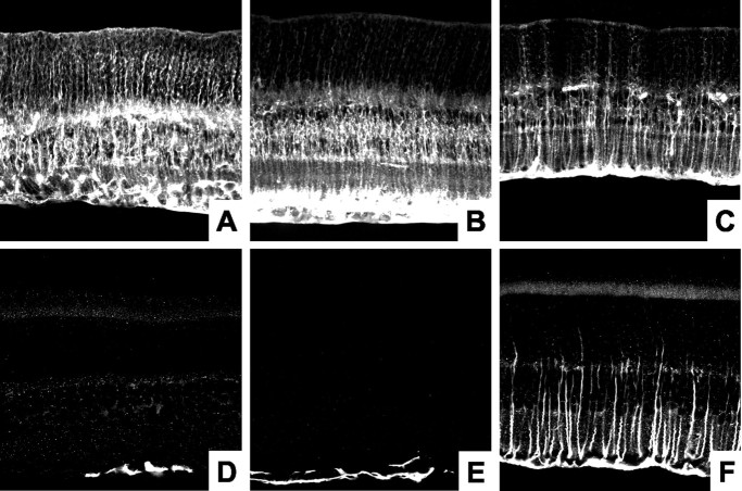
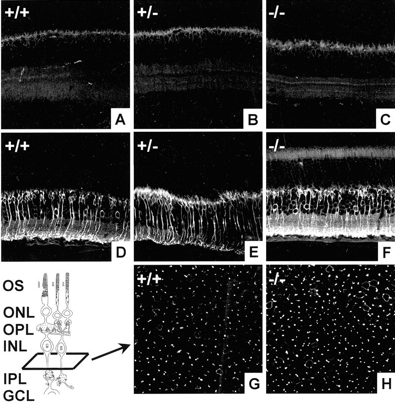
BETA2/NeuroD1 is a basic helix-loop-helix transcription factor that is expressed widely throughout the developing nervous system. Previous studies have shown that BETA2/NeuroD1 influences the fate of retinal cells in culture. To analyze the effect of BETA2/NeuroD1 on the structure and function of the retina, we examined a line of BETA2/NeuroD1 knock-out mice that survives until adulthood. At 2-3 months of age, homozygous null mice showed a 50% reduction in rod-driven electroretinograms (ERGs) and a 65% reduction in cone-driven ERGs. ERGs measured from knock-out mice that were >9 months of age were undetectable. At 2-3 months, the number of photoreceptors in the outer nuclear layer was reduced by 50%. In addition, electron microscopy showed that the surviving photoreceptors had shortened outer segments. The number of cones labeled by peanut agglutinin was decreased 50-60%. By 18 months, retinas from null mice were completely devoid of photoreceptors. There appeared to be few changes in the inner retina, although BETA2/NeuroD1 is expressed in this area. Terminal deoxynucleotidyl transferase-mediated biotinylated UTP nick end labeling staining revealed a dramatic increase in cell death, peaking at approximately postnatal day 3 and continuing into adulthood. No defects in cell birth were detected using bromodeoxyuridine staining. Our results reveal that BETA2/NeuroD1 not only plays an important role in terminal differentiation of photoreceptors but also serves as a potential survival factor. Loss of BETA2/NeuroD1 results in an age-related degeneration of both rods and cones.
Figures
Similar articles
-
Ophthalmological phenotype associated with homozygous null mutation in the NEUROD1 gene.Mol Vis. 2015 Feb 5;21:124-30. eCollection 2015. Mol Vis. 2015. PMID: 25684977 Free PMC article.
-
NeuroD1 is required for survival of photoreceptors but not pinealocytes: results from targeted gene deletion studies.J Neurochem. 2012 Oct;123(1):44-59. doi: 10.1111/j.1471-4159.2012.07870.x. Epub 2012 Aug 16. J Neurochem. 2012. PMID: 22784109 Free PMC article.
-
Detailed histopathologic characterization of the retinopathy, globe enlarged (rge) chick phenotype.Mol Vis. 2005 Jan 13;11:11-27. Mol Vis. 2005. PMID: 15660021
-
Disease model: photoreceptor degenerations.Trends Mol Med. 2001 Mar;7(3):133-5. doi: 10.1016/s1471-4914(00)01902-x. Trends Mol Med. 2001. PMID: 11286785 Review.
-
Vertebrate photoreceptor cell development and disease.Trends Cell Biol. 1998 Sep;8(9):353-8. doi: 10.1016/s0962-8924(98)01341-5. Trends Cell Biol. 1998. PMID: 9728396 Review.
Cited by
-
Dlic1 deficiency impairs ciliogenesis of photoreceptors by destabilizing dynein.Cell Res. 2013 Jun;23(6):835-50. doi: 10.1038/cr.2013.59. Epub 2013 Apr 30. Cell Res. 2013. PMID: 23628724 Free PMC article.
-
Neurogenin and NeuroD direct transcriptional targets and their regulatory enhancers.EMBO J. 2007 Dec 12;26(24):5093-108. doi: 10.1038/sj.emboj.7601923. Epub 2007 Nov 15. EMBO J. 2007. PMID: 18007592 Free PMC article.
-
Identification of molecular markers of bipolar cells in the murine retina.J Comp Neurol. 2008 Apr 10;507(5):1795-810. doi: 10.1002/cne.21639. J Comp Neurol. 2008. PMID: 18260140 Free PMC article.
-
Reprogramming progeny cells of embryonic RPE to produce photoreceptors: development of advanced photoreceptor traits under the induction of neuroD.Invest Ophthalmol Vis Sci. 2008 Sep;49(9):4145-53. doi: 10.1167/iovs.07-1380. Epub 2008 May 9. Invest Ophthalmol Vis Sci. 2008. PMID: 18469196 Free PMC article.
-
A role of ath5 in inducing neuroD and the photoreceptor pathway.J Neurosci. 2004 Aug 11;24(32):7150-8. doi: 10.1523/JNEUROSCI.2266-04.2004. J Neurosci. 2004. PMID: 15306648 Free PMC article.
References
-
- Acharya HR, Dooley CM, Thoreson WB, Ahmad I. cDNA cloning and expression analysis of NeuroD mRNA in human retina. Biochem Biophys Res Commun. 1997;233:459–463. - PubMed
-
- Ahmad I, Acharya HR, Rogers JA, Shibata A, Smithgall TE, Dooley CM. The role of NeuroD as a differentiation factor in the mammalian retina. J Mol Neurosci. 1998;11:165–178. - PubMed
-
- Bessant DA, Payne AM, Mitton KP, Wang QL, Swain PK, Plant C, Bird AC, Zack DJ, Swaroop A, Bhattacharya SS. A mutation in NRL is associated with autosomal dominant retinitis pigmentosa. Nat Genet. 1999;21:355–356. - PubMed
-
- Blanks JC, Johnson LV. Selective lectin binding of the developing mouse retina. J Comp Neurol. 1983;221:31–41. - PubMed
-
- Brown NL, Kanekar S, Vetter ML, Tucker PK, Gemza DL, Glaser T. Math5 encodes a murine basic helix-loop-helix transcription factor expressed during early stages of retinal neurogenesis. Development. 1998;125:4821–4833. - PubMed
Publication types
MeSH terms
Substances
Grants and funding
LinkOut - more resources
Full Text Sources
Molecular Biology Databases