. 2003 Jan 15;23(2):470-80.
doi: 10.1523/JNEUROSCI.23-02-00470.2003.

Light stimulates a transducin-independent increase of cytoplasmic Ca2+ and suppression of current in cones from the zebrafish mutant nof

Affiliations

Light stimulates a transducin-independent increase of cytoplasmic Ca2+ and suppression of current in cones from the zebrafish mutant nof

Susan E Brockerhoff et al. J Neurosci. .

Abstract

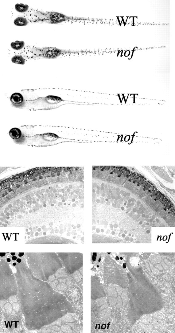
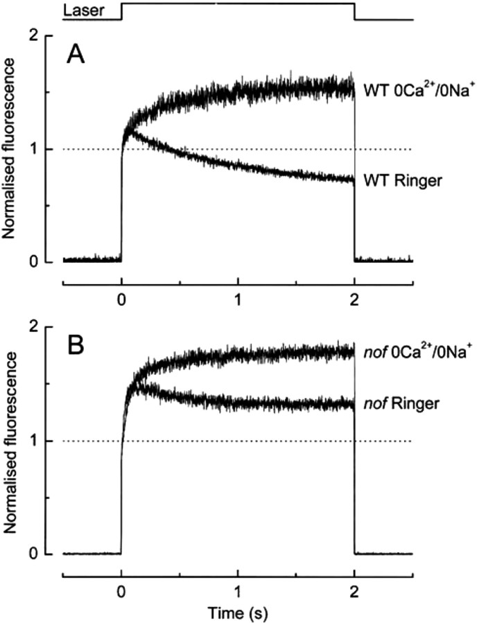
Transducins couple visual pigments to cGMP hydrolysis, the only recognized phototransduction pathway in vertebrate photoreceptors. Here we describe a zebrafish mutant, no optokinetic response f(w21) (nof), with a nonsense mutation in the gene encoding the alpha subunit of cone transducin. Retinal morphology and levels of phototransduction enzymes are normal in nof retinas, but cone transducin is undetectable. Dark current in nof cones is also normal, but it is insensitive to moderate intensity light. The nof cones do respond, however, to bright light. These responses are produced by a light-stimulated, but transducin-independent, release of Ca2+ into the cone cytoplasm. Thus, in addition to stimulating transducin, light also independently induces release of Ca2+ into the photoreceptor cytoplasm.


Figures

Fig. 1.
Fig. 1.
Identification of the Tcα mutation innof. A, The nof mutation changes nucleotide 154 from a C to a T to introduce a stop codon (TAG) at amino acid position 52. B, Alignment of zebrafish Tcα with bovine Tcα. Nonconservative changes are indicated inbold.
Fig. 2.
Fig. 2.
Tcα mRNA expression in larval and adult WT zebrafish. AE, Antisense mRNA expression; F, sense control. Age of samples is between 52 and 56 hpf (A), between 70 and 74 hpf (B, C), between 79 nd 83 hpf (D), and adult (E,F). Refer to Results for a detailed description of the staining pattern. p, Pineal;n, nasal; t, temporal; d, dorsal; v, ventral; y, yolk. The dark staining of the yolk is nonspecific and is frequently seen with many other probes (Westerfield, 1995).
Fig. 3.
Fig. 3.
Tcα mRNA and protein expression innof versus OKR+ zebrafish. A, In situ hybridization of nof and OKR+ larvae at 5 dpf shows that Tcα mRNA is dramatically reduced in thenof mutant. B, Western blot analysis of eye homogenates using affinity-purified Tcα polyclonal antibody. Tcα protein is undetectable in the nof mutant.C, Immunocytochemical analysis of OKR+ andnof retinas at 2 months using affinity-purified Tcα antibody. ros, Rod outer segments; dc, double cone; ls, long single cone; ss, short single cone; opl, outer plexiform layer;inl, inner nuclear layer.
Fig. 4.
Fig. 4.
Normal morphology of the nofmutant. A, Dorsal (top) and lateral (bottom) view of nof and OKR+ larvae at 5 dpf. The nof mutant appears morphologically normal. Typical length of a 5 dpf is ∼6 mm. B,C, The retina of nof appears normal. Light (B) and transmission electron microscopy (C) of nof and OKR+ retinas at 5–6 dpf are shown. Typical diameter of the eye at 5 dpf is ∼300 μm. Scale bar (shown in C): 1 μm.PRL, Photoreceptor layer; ONL, outer nuclear layer; IPL, inner plexiform layer;GCL, ganglion cell layer; L, lens.
Fig. 5.
Fig. 5.
A, zpr1 staining of 2-month-old OKR+ and nof retinas. ros, Rod outer segments; dcis, double cone inner segments.B, PDE activity appears normal in the nofmutant. Measurements of basal and maximal PDE activity in homogenates of eyes from 5–7 dpf OKR+ and nof larvae are shown.C, Western blot analysis of homogenates of eyes from 5–7 dpf OKR+ and nof larvae. Lanes 1 and2 were labeled with an antibody that recognizes zebrafish rod and cone Tα subunits. Each lanecorresponds to 5–10 larval eyes.
Fig. 6.
Fig. 6.
Photoreceptor responses of the nofmutant. A, Current recordings from an OKR+ L cone in response to 10 msec flashes of 590 nm light. The dimmest flash had a strength of 630 photons per square micrometer, and each successive flash was twice as bright. B, Response from anof cone to a 10 msec flash of 44100 photons per square micrometer. For comparison the response of the OKR+ L cone from B to a flash 70× dimmer is shown. C, Comparison of the flash sensitivity of rods and cones from OKR+ (control) andnof fish. In contrast to cones, nof rods have normal sensitivity. Error bars are SE from recordings from 4nof rods, 5 OKR+ rods, 73 nof cones, and 6 OKR+ cones. Open circle, OKR+ rod; closed circle, nof rod; open diamond, OKR+ cone;closed diamond, nof cone.
Fig. 7.
Fig. 7.
Small, Ca2+-sensitive electrical responses are detected in nof cones during light steps. A, Shown is a family of step responses of 2 sec duration from an OKR+ L cone. The dimmest step had an intensity of 1575 photons per square micrometer per second, and each successive step was twice as bright. B, Comparison of step response from an OKR+ cone with the response of a nof cone to a light step 400× brighter. C, Average response of 26nof cones to a 2 sec light step of intensity 1.3 × 10⁷ photons per square micrometer per second. The averaged amplitude is significantly smaller than that obtained from the majority of individual cells because this average includes everynof cone from which responses to this step were measured. Some of the cells included in the average did not appear to respond. D, Comparison of the maximum amplitudes ofnof cone responses with and without BAPTA loading. BAPTA dramatically reduced the response from nof cones. Theinset shows the averaged responses from 2 sec stimuli. Calibration, 0.2 pA. The step intensity was 6.5 × 10⁶ photons per square micrometer per second, and in all cases 590 nm light stimuli were used.
Fig. 8.
Fig. 8.
Change in free Ca2+ in cones from OKR+ and OKR− zebrafish cones. Fluorescence was recorded from zebrafish cones preloaded with fluo-4, a reporter of free Ca2+ concentration. Cones containing the dye were stimulated for 2 sec with intense laser light (see Materials and Methods). Traces are means from 13–25 cells, individually normalized to the mean value of the fluorescence recorded during the first 20 msec of laser illumination. A, Cones from OKR+ (WT) animals in Ringer's (bottom trace) produce a small increase in fluorescence followed by a decrease to a level substantially below the initial value. In 0Ca2+/0Na+ solution (top trace), the fluorescence increases, but there is no subsequent decline. B, Stimulation of nof cones in Ringer's (bottom trace) produces a large increase in fluorescence followed by a slight decrease. Note that the final value of fluorescence remains substantially higher than the initial value, unlike in OKR+ cones. In 0Ca2+/0Na+ solution (top trace), the fluorescence from nof cones increases with no subsequent decline, similar to 0Ca2+/0Na+ responses from OKR+ cones.

References

    1. Arslan P, Di Virgilio F, Beltrame M, Tsien RY, Pozzan T. Cytosolic Ca2+ homeostasis in Ehrlich and Yoshida carcinomas. A new, membrane-permeant chelator of heavy metals reveals that these ascites tumor cell lines have normal cytosolic free Ca2+. J Biol Chem. 1985;260:2719–2727. - PubMed
    1. Barthel LK, Raymond PA. In situ hybridization studies of retinal neurons. Methods Enzymol. 2000;316:579–590. - PubMed
    1. Branchek T. The development of photoreceptors in the zebrafish, brachydanio rerio. II. Function. J Comp Neurol. 1984;224:116–122. - PubMed
    1. Brockerhoff SE, Hurley JB, Janssen-Bienhold U, Neuhauss CF, Driever W, Dowling JE. A behavioral screen for isolating zebrafish mutants with visual system defects. Proc Natl Acad Sci USA. 1995;92:10545–10549. - PMC - PubMed
    1. Brockerhoff SE, Hurley JB, Niemi GA, Dowling JE. A new form of inherited red-blindness identified in zebrafish. J Neurosci. 1997;20:1–8. - PMC - PubMed

Publication types

MeSH terms