. 2003 Jan 15;23(2):493-502.
doi: 10.1523/JNEUROSCI.23-02-00493.2003.

Aberrant activation of focal adhesion proteins mediates fibrillar amyloid beta-induced neuronal dystrophy

Affiliations

Aberrant activation of focal adhesion proteins mediates fibrillar amyloid beta-induced neuronal dystrophy

Elizabeth A Grace et al. J Neurosci. .

Abstract

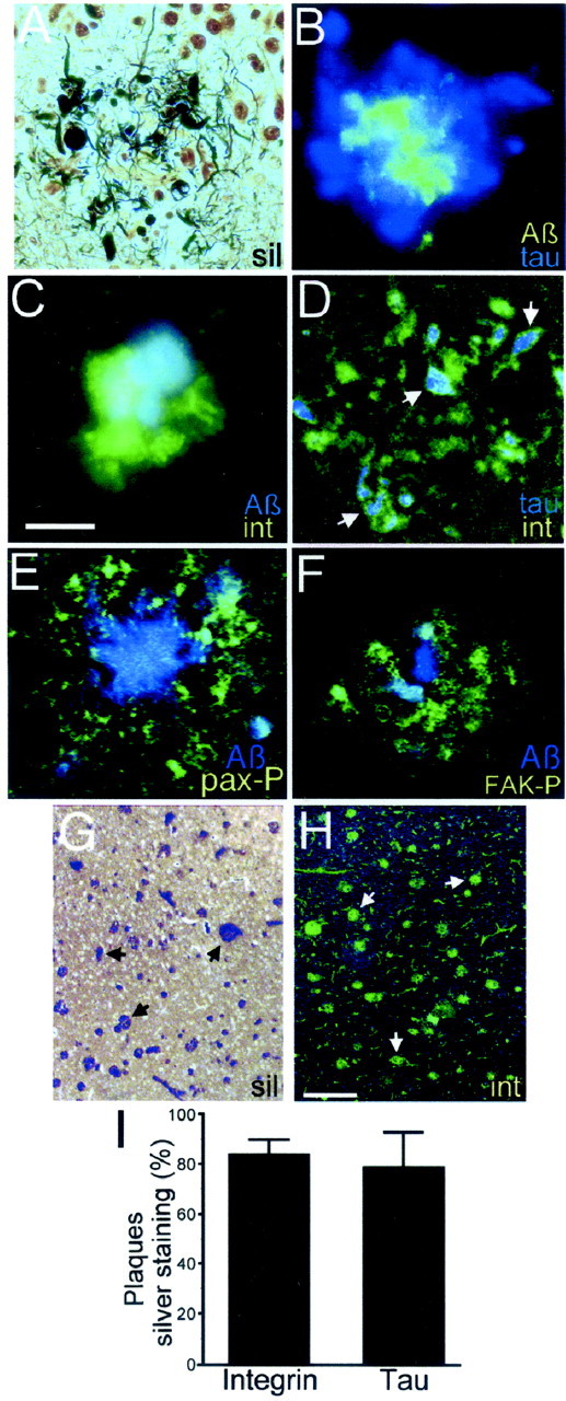
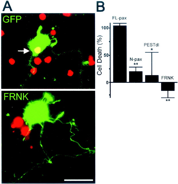
Neuronal dystrophy is a pathological hallmark of Alzheimer's disease (AD) that is not observed in other neurodegenerative disorders that lack amyloid deposition. Treatment of cortical neurons with fibrillar amyloid beta (Abeta) peptides induces progressive neuritic dystrophy accompanied by a marked loss of synaptophysin immunoreactivity (Grace et al., 2002). Here, we report that fibrillar Abeta-induced neuronal dystrophy is mediated by the activation of focal adhesion (FA) proteins and the formation of aberrant FA structures adjacent to Abeta deposits. In the AD brain, activated FA proteins are observed associated with the majority of senile plaques. Clustered integrin receptors and activated paxillin (phosphorylated at Tyr-31) and focal adhesion kinase (phosphorylated at Tyr-297) are mainly detected in dystrophic neurites surrounding Abeta plaque cores, where they colocalize with hyperphosphorylated tau. Deletion experiments demonstrated that the presence of the LIM domains in the paxillin C terminus and the recruitment of the protein-Tyr phosphatase (PTP)-PEST to the FA complex are required for Abeta-induced neuronal dystrophy. Therefore, both paxillin and PTP-PEST appear to be critical elements in the generation of the dystrophic response. Paxillin is a scaffolding protein to which other FA proteins bind, leading to the formation of the FA contact and initiation of signaling cascades. PTP-PEST plays a key role in the dynamic regulation of focal adhesion contacts in response to extracellular cues. Thus, in the AD brain, fibrillar Abeta may induce neuronal dystrophy by triggering a maladaptive plastic response mediated by FA protein activation and tau hyperphosphorylation.


Figures

Fig. 1.
Fig. 1.
Fibrillar Aβ induces neuronal dystrophy, Tyr phosphorylation, and paxillin activation in cortical neurons. Aβ was added at day 5, and the cultures were processed at day 7 in culture.A, Neurites appear smooth and healthy incontrol cultures. Fibrillar Aβ-induced aberrant neurite morphology, including decreased caliber and acute angles and loops (Aβ, arrows). Neurons were immunolabeled with anti-tubulin class III antibody. Scale bar, 10 μm.B, Western blot of whole-cell homogenates developed with anti-phospho-Tyr antibody 4G10. Note the increase in phospho-Tyr in several bands after Aβ treatment (arrowheads) and decreased Tyr phosphorylation in a band of ∼180 kDa (small arrow). Con, Control. C, Homogenates were immunoprecipitated (IP) with anti-paxillin (α-pax) antibody and immunostained with 4G10. Note the increase in paxillin Tyr phosphorylation in Aβ-treated neurons (Aβ). Immunoprecipitates with nonimmune (NI) serum were negative. Con, Control; WB, Western blot. D, Paxillin Tyr phosphorylation was quantified by densitometry in vehicle-treated samples (Con) and normalized as 100%. Note the significant increase (170 ± 30%) induced by fibrillar Aβ (Aβ). The membranes were reprobed for paxillin to confirm equal loading. Values are mean ± SEM;n = 4 independent experiments; *p < 0.05. E, Western blot analysis of whole-cell homogenates and cytoskeletal extracts. The blots were developed with anti-paxillin antibody (pax). Note the increase in paxillin (pax) in the cytoskeletal fraction (Cytosk) of Aβ-treated neurons (Aβ). Total paxillin levels in whole-cell homogenates did not change. Similar tubulin levels (tub) confirmed equal loading. Con, Control. F, Densitometric quantification of paxillin in the cytoskeleton revealed a 680 ± 200% increase in Aβ-treated neurons grown on poly-l-lysine and a 210 ± 30% increase in control (Con) neurons grown on laminin (Lam).n = 9 independent experiments; *p < 0.05 relative to control by Student'st test.
Fig. 2.
Fig. 2.
Fibrillar Aβ induces FA-like structures in dystrophic neurons. Cortical neurons were treated with fibrillar Aβ at day 5, fixed at day 7, and stained with phalloidin-Texas Red, anti-Aβ, anti-paxillin (pax), and anti-integrin (int) antibodies. FAs were identified by colocalization of paxillin or integrin clusters with microfilaments.A–D, FA are absent in neurons grown on poly-l-lysine (control, PLL). Neuronal processes exhibit homogeneous distribution of paxillin and integrin, whereas microfilaments (MF, red) are primarily localized in growth cones (A, C, arrows). E, F, Neurons grown on laminin (control, Lam) exhibit paxillin and integrin clusters (arrows), associated with microfilaments periodically localized along the processes.G–J, Aβ-treated neurons on poly-l-lysine [Aβ (PLL)] develop FA-like structures proximal to Aβ fibrils (blue) that include clusters of paxillin and integrin (arrows). Microfilaments (MF, red) protruding from FA-like structures are evident in G and H (arrowheads). In some cases, growth cone filopodia appear to reverse orientation and extend toward Aβ deposits (I, J, arrowheads).K–N, Phospho-Tyr immunoreactivity (Tyr-P, green) colocalizes with paxillin (blue) and microfilaments (MF, red) in dystrophic processes (arrows). Scale bar: (in F)A–N, 5 μm. O, Quantification of integrin and paxillin clustering. Integrin receptor clustering increased 2.0 ± 0.2-fold on a laminin substrate and 1.8 ± 0.2-fold in neurons grown on poly-l-lysine after Aβ treatment. Paxillin clustering increased 3.0 ± 0.3-fold on a laminin substrate and 2.5 ± 0.3-fold in neurons grown on poly-l-lysine after Aβ treatment. Values are mean ± SEM; n = 3 independent experiments; >100 FA contacts were scored per condition; *p < 0.05 relative to control by ANOVA followed by the Student–Newman–Keulspost hoc test.
Fig. 3.
Fig. 3.
Expression of FA proteins in Alzheimer's brain. Paraffin-embedded brain sections of AD and age-matched control specimens were silver-stained or immunolabeled. A, In AD brains, silver staining revealed dystrophic neurites (black) in senile plaques, surrounding the core of Aβ (pale yellow). B, Immunofluorescence shows the Aβ core of the plaque (green) surrounded by dystrophic neurites immunostained with antibody PHF-1, which recognizes hyperphosphorylated tau (blue). C, Integrin receptor immunoreactivity in a senile plaque (green) surrounding the Aβ core (blue). D, Hyperphosphorylated tau (blue) and integrin receptors (green) colocalize in dystrophic neurites and cell bodies in a senile plaque (arrows).E, Phosphorylated paxillin (pax-P,green) in dystrophic neurites and cell bodies in a senile plaque. The plaque core is stained with anti-Aβ antibody (Aβ, blue). F, Dystrophic neurites in a senile plaque immunostained with anti-phosphorylated FAK antibody (FAK-P, green) surrounding the Aβ core (blue). Adjacent brain sections silver-stained (G) and immunostained with anti-integrin antibody (int,H) show similar plaque density.Arrows denote individual plaques. I, Quantification of integrin- and hyperphosphorylated tau-positive plaques shows that 84 ± 6% of silver-stained plaques were positive for integrin immunoreactivity, and 79 ± 14% were positive for hyperphosphorylated tau. Ten to 20 microscopic fields were analyzed in adjacent sections of four AD brain cases. At least 50 plaques were scored per silver-stained section. Scale bars:A–F, 50 μm; G, H, 250 μm.
Fig. 4.
Fig. 4.
Paxillin LIM domains are required for Aβ-induced neuronal dystrophy. A, Cortical neurons were transfected with GFP or GFP-paxillin expression vectors containing full-length paxillin (FL-pax) or deletions of the LIM domains (N-pax) or the LIM domains (LIM-pax), respectively. B, Cortical neurons were transfected at day 5 and treated with fibrillar Aβ. The number of dystrophic neurons was quantified 24 hr later and expressed as a percentage of the number of Aβ-induced dystrophic neurons expressing GFP (100%). Note the significant inhibition of neuronal dystrophy in neurons expressingN-pax (28.6 ± 15.1%). FL-pax and LIM-pax were not significantly different from GFP (FL-pax, 108 ± 12%; LIM-pax, 90.4 ± 21.9%). Values are the mean ± SEM; n = 4 independent experiments; 250 neurons were scored per experiment; *p < 0.05 relative to GFP-transfected cells by ANOVA. C–E, Neurons expressing the indicated GFP-tagged paxillin proteins were immunostained with anti-Aβ antibody (blue) and phalloidin-Texas Red. Note the formation of FA-like structures in neurons expressing FL-pax, N-pax, and LIM-pax (arrows).
Fig. 5.
Fig. 5.
PTP-PEST binding to paxillin is required for Aβ-induced neuronal dystrophy. A, Aβ-induced neuronal dystrophy was assessed in neurons transfected with paxillin constructs bearing Cys-to-Ala point mutations, which disrupt the LIM domain tertiary structure, with FRNK, an FAK dominant negative construct, or with PESTdl, a PTP-PEST deletion construct.B, Mutations C470A, C467/470A, and C523A in paxillin, which prevent PTP-PEST binding and integrin association, significantly reduce Aβ-induced neuronal dystrophy (C470A, 62.6 ± 1.3%; C467/C470A, 21.2 ± 4.4%;C523A, 39.9 ± 18.5%). The mutationC467A, which also prevents PTP-PEST binding and integrin association, has no effect on Aβ-induced neuronal dystrophy (112.2 ± 36.1). Expression of PESTdl, which prevents PTP-PEST binding to paxillin, completely prevents neuronal dystrophy. Expression of FRNK, which contains the FAK focal adhesion targeting domain but lacks the kinase domain, has no effect on Aβ-induced neuronal dystrophy. The number of dystrophic neurons was quantified 24 hr after transfection and expressed as a percentage of the number of Aβ-induced dystrophic neurons expressing GFP (100%). Values are mean ± SEM; n = 3–7 individual experiments; 250 neurons were scored per condition in each experiment; *p < 0.05; **p < 0.01 relative to control (GFP) by ANOVA followed by the Student–Newman–Keuls post hoc test.
Fig. 6.
Fig. 6.
PTP-PEST and FAK activity are involved in Aβ-induced neuronal death. A, Aβ treatment induces cell death in neurons expressing GFP (top panel, greenfluorescence). Nonviable cells show process retraction and disintegration and positive nuclear staining for propidium iodide (arrow). Neurons expressing FRNK (bottom panel, green fluorescence) became dystrophic but remained viable. Note the dystrophic appearance of neuritic processes and the absence of propidium nuclear staining in the neuron expressing FRNK.B, Quantification of cell death in transfected neurons treated with Aβ. A significant reduction in Aβ-induced neuronal death is observed in neurons expressing N-pax, which lacks paxillin LIM domains (20.4 ± 8%), andPESTdl, which lacks the paxillin-binding domain of PTP-PEST (13.3 ± 41.7%). FRNK completely prevents neuronal death (−15.3 ± 13%). Cell death is expressed as a percentage of Aβ-induced propidium-positive neurons transfected with GFP (100%). Values are mean ± SEM; n = 3 independent experiments; >200 neurons were scored per condition in each individual experiment; *p < 0.05; **p < 0.01 relative to control (GFP) by ANOVA followed by the Student–Newman–Keuls post hoc test. Cortical neurons were transfected at day 5, treated with fibrillar Aβ for 2 d, and processed for analysis. Before fixation, the nuclei of dead cells were labeled with propidium iodide.
Fig. 7.
Fig. 7.
Model of the FA pathways involved in Aβ-induced neuronal dystrophy. Fibrillar Aβ binds to and induces the clustering of the integrin receptors, leading to the activation of paxillin and FAK and their translocation to the nascent FA complex. Paxillin binds to vinculin, which promotes microfilament stabilization at the FA site. PTP-PEST binds to paxillin, leading to dephosphorylation of several FA proteins, which prevents the stabilization of the FA contact, allowing the neuron to continuously respond to fibrillar Aβ stimuli. Alternatively, APP binds to Aβ fibrils, bringing them in contact with integrins, activating FA signaling through FE65, which binds to the C terminus of APP and associates with c-abl, which in turn binds and phosphorylates paxillin, or both. A pathway involving fyn, which is downstream of PTP-PEST, promotes GSK3β activity, whereas the interaction of cbl with c-abl increases CDK5 activity (Zukerberg et al., 2000). Both CDK5 and GSK3β hyperphosphorylate tau, leading to microtubular destabilization and neuronal dystrophy. An alternative pathway involving FAK activity leads to neuronal cell death but not neuronal dystrophy. Paxillin contains four SH2-binding domains (red), five Leu-rich LD domains (light blue), one Pro-rich SH3-binding domain (green) and four LIM domains (purple). MF, microfilaments;hyperphosp., hyperphosphorylated; P, phosphate groups.

Similar articles

Cited by

References

    1. Akiyama H, Kawamata T, Dedhar S, McGeer PL. Immunohistochemical localization of vitronectin, its receptor and beta-3 integrin in Alzheimer brain tissue. J Neuroimmunol. 1991;32:19–28. - PubMed
    1. Angers-Loustau A, Cote JF, Charest A, Dowbenko D, Spencer S, Lasky LA, Tremblay ML. Protein tyrosine phosphatase-PEST regulates focal adhesion disassembly, migration, and cytokinesis in fibroblasts. J Cell Biol. 1999;144:1019–1031. - PMC - PubMed
    1. Aplin AE, Howe A, Alahari SK, Juliano RL. Signal transduction and signal modulation by cell adhesion receptors: the role of integrins, cadherins, immunoglobulin-cell adhesion molecules, and selectins. Pharmacol Rev. 1998;50:197–263. - PubMed
    1. Baumann K, Mandelkow EM, Biernat J, Piwnica-Worms H, Mandelkow E. Abnormal Alzheimer-like phosphorylation of tau-protein by cyclin-dependent kinases cdk2 and cdk5. FEBS Lett. 1993;336:417–424. - PubMed
    1. Benzing WC, Mufson EJ, Armstrong DM. Alzheimer's disease-like dystrophic neurites characteristically associated with senile plaques are not found within other neurodegenerative diseases unless amyloid beta-protein deposition is present. Brain Res. 1993;606:10–18. - PubMed

Publication types

MeSH terms

LinkOut - more resources