. 2003 Feb 4;100(3):886-91.
doi: 10.1073/pnas.0237311100. Epub 2003 Jan 17.

A photochemical approach to the lipid accessibility of engineered cysteinyl residues

Affiliations

A photochemical approach to the lipid accessibility of engineered cysteinyl residues

Jing Li et al. Proc Natl Acad Sci U S A. .

Abstract

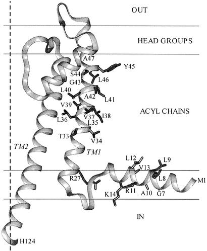
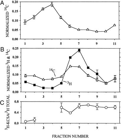
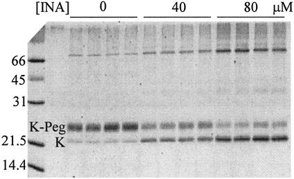
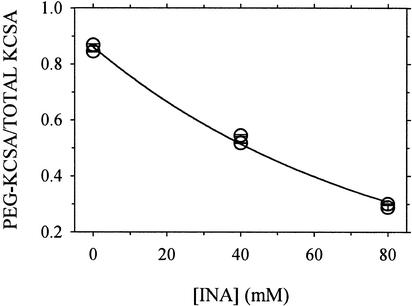
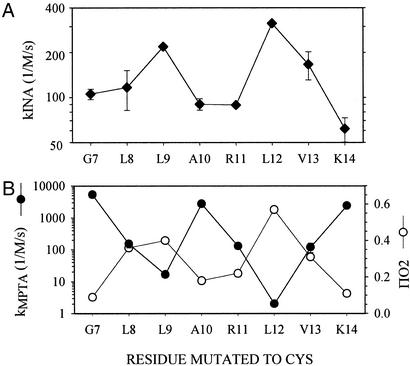
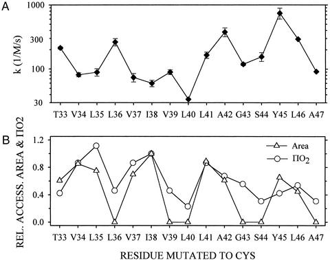
Ordinary electrophilic reagents react too slowly in a nonpolar environment to be useful for the determination of the accessibility to lipid of continuous stretches of residues mutated to cysteine. By contrast, photoactivated 5-iodonaphthyl-1-azide (INA) reacted readily with 2-mercaptoethanol and dodecanethiol in nonpolar solvents and in liposomes. Continuous stretches of residues in the amphipathic N-terminal helix and first transmembrane helix of the bacterial potassium channel KcsA were replaced with cysteine, and the mutants were expressed in Escherichia coli and isolated in inner membranes. These membranes were dissolved in detergent and reconstituted into asolectin liposomes incorporating INA. The extent of light-induced reaction of INA with each cysteine was assayed by subsequent reaction with the gel-shifting, SH-specific methoxy-polyethylene glycol-2-pyridine disulfide. The pattern of apparent second-order rate constants for the photoreactions of eight substituted cysteines in the N-terminal helix conformed to other measures of lipid exposure. The pattern of the rate constants for the photoreactions of 15 cysteines in the first transmembrane helix had peaks every third residue, which partly conformed to other measures of lipid exposure.


Figures

Figure 1
Figure 1
Structure of the KcsA monomer and its location in the phospholipid bilayer. The coordinates of residues 22–124 were from the crystal structure 1K4C (26). Residues 1–17 were configured in INSIGHT II as an ideal α-helix, which was joined to residues 22–124 by residues 18–21 in a loop. We generated a loop using the loop module in the modeler program and the modeler objective function (28). We manipulated the locations of the core residues 22–124 and the N-terminal helix relative to the 30-Å-wide acyl-chain domain and the 7.5-Å-wide head-group domains so as to place the R27 guanidinium in the head-group domain and the lipid-facing residues of the N-terminal helix (11, 27) in contact with the acyl chains. In addition to R27, the side chains of the residues probed in this article are shown. The fourfold axis of the tetramer, which is also the axis of the channel, is shown as a broken line.
Figure 2
Figure 2
Association of KcsA and phospholipid in reconstituted liposomes. Liposomes consisting of asolectin and 14C-phospholipid, suspended in KCa buffer, were added to DM/KCa buffer (A) or to 3H-labeled KcsA in DM/KCa buffer (B) and reconstituted by dilution. The concentrations of asolectin, protein, and DM and the reconstitution conditions were the same as those given in Materials and Methods. The mixture of asolectin and 14C-phospholipid was made by mixing 150 μl of 20 mg of asolectin per ml of CHCl3 and 300 nCi (1 Ci = 37 GBq) of 14C-dipalmitoyl phosphatidylcholine (25 nCi/μl of toluene/ethanol, 1:1). 3H-labeled KcsA was made by reacting E. coli inner membrane containing 25 μg of KcsA mutant K14C and 162 μg of total protein with 10 mM DTT (pH 8.0) for 60 min. This suspension was sedimented, and the pellet was suspended in 20 mM Hepes (pH 7.0)/1 mM EDTA, sedimented, and suspended again to lower the DTT concentration. The reduced protein was reacted with 4.4 μM 3H-NEM/90 μM NEM for 30 min and sedimented and suspended three times to lower the free 3H-NEM concentration. In duplicate, 200 μl of reconstituted liposomes containing 14C-phospholipid (A) and 200 μl of reconstituted liposomes containing 3H-KcsA and 14C-phospholipid (B) were layered over 4 ml of a linear gradient from 5–31% (wt/wt) sucrose in 10 mM Hepes (pH 7.0), and sedimented in a Beckman SW60 rotor at 60,000 rpm for 13 h at 4°C. The sucrose concentration and linearity of the gradient were determined in parallel tubes with an Abbe refractometer (Zeiss). Ten 400-μl fractions were taken from the top (fraction 1 is topmost), and the volume of the remaining 11th fraction, which was <400 μl, was determined. Forty-microliter aliquots of each fraction were mixed with 5 ml of scintillation fluid and counted with a double-label protocol. The average radioactivity (and average error), normalized by the total recovered radioactivity, is plotted versus the fraction number. (C) The part of the total 3H radioactivity that was associated with KcsA in each fraction in B was determined by diluting the remaining 360 μl of each fraction (less in fraction 11) with 0.5 ml of 1% DM/100 mM NaCl/50 mM NaPi (pH 8.0), stirring with 50 μl of NiNTA-agarose (1:1) for 60 min, washing the gel, and eluting the bound KcsA in two 50-μl aliquots of 5 mM EDTA/LSB (conditions as in Materials and Methods). The combined eluted 3H radioactivity was determined and is plotted versus the sucrose-density-gradient B fraction number normalized by the input to the NiNTA-agarose. There was too little 3H-labeled protein in sucrose-density-gradient fractions 3 and 4 to determine its binding to NiNTA-agarose.
Figure 3
Figure 3
Electrophoresis of KcsA reacted with INA and PegSSP. The reaction and SDS/PAGE conditions are given in Materials and Methods. The final concentrations of INA averaged over the volume of the liposome suspension are at the top of the gel. For each INA concentration, duplicate lanes were run of each of duplicate reaction mixtures. The positions of the KcsA monomer (K) and of the KcsA-PegSSP adduct (K-Peg) are indicated. The molecular masses of protein standards are in kDa.
Figure 4
Figure 4
The fraction of T33C that reacted with PegSSP after photoreaction with INA. The data are from the gel shown in Fig. 3. The integrated densities of the KcsA monomer band and of the KcsA-PegSSP adduct band were determined and added, and the fraction (y) of the total in the KscA-PegSSP adduct band was calculated. The means of the duplicate lanes (and average errors) for each of the duplicate reactions are shown. The errors in all cases were smaller than the symbol. A nonlinear least-squares fit to the data of the equation y = ae−b[INA] yielded b, which was divided by the time of photolysis, 60 s, to yield an apparent second-order rate constant, kINA (M−1⋅s−1).
Figure 5
Figure 5
Rate constants for the photoreaction of INA with Cys-substitution mutants in the N-terminal amphipathic helix. (A) Apparent second-order rate constants were obtained as described in the legend of Fig. 4. Means and errors (most are within the symbols) in the fits of duplicate reaction mixtures are shown. (B) For comparison, we show the rate constants for the reactions of the hydrophilic 4-maleimidophenyltrimethylammonium (kMPTA) with the Cys mutants (11) and the O2-accessibility parameter (ΠO2) from the quenching by O2 of spin-labeled Cys mutants (27). Note the rate constants are on logarithmic scales and the quenching parameter is on a linear scale.
Figure 6
Figure 6
Rate constants for the photoreaction of INA with Cys-substitution mutants in TM1. (A) Apparent second-order rate constants were obtained as described in the legend of Fig. 4. Means and errors in the fits of duplicate or quadruplicate reaction mixtures are shown. (B) For comparison, we show ΠO2 (29) and the molecular surface area of the Cys sulfur atom, which is in each case relative to these values for I38C (see Materials and Methods).

Similar articles

Cited by

References

    1. Kaback H R, Sahin-Toth M, Weinglass A B. Nat Rev Mol Cell Biol. 2001;2:610–620. - PubMed
    1. Hubbell W L, Cafiso D S, Altenbach C. Nat Struct Biol. 2000;7:735–739. - PubMed
    1. Karlin A, Akabas M H. Methods Enzymol. 1998;293:123–145. - PubMed
    1. Falke J J, Hazelbauer G L. Trends Biochem Sci. 2001;26:257–265. - PMC - PubMed
    1. Zhang H, Karlin A. Biochemistry. 1998;37:7952–7964. - PubMed

Publication types

MeSH terms

LinkOut - more resources