Comparative Study
. 2002;3(12):RESEARCH0068.
doi: 10.1186/gb-2002-3-12-research0068. Epub 2002 Nov 12.

Membrane-bound progesterone receptors contain a cytochrome b5-like ligand-binding domain

Affiliations
Comparative Study

Membrane-bound progesterone receptors contain a cytochrome b5-like ligand-binding domain

William Mifsud et al. Genome Biol. 2002.

Abstract

Background: Membrane-associated progesterone receptors (MAPRs) are thought to mediate a number of rapid cellular effects not involving changes in gene expression. They do not show sequence similarity to any of the classical steroid receptors. We were interested in identifying distant homologs of MAPR better to understand their biological roles.

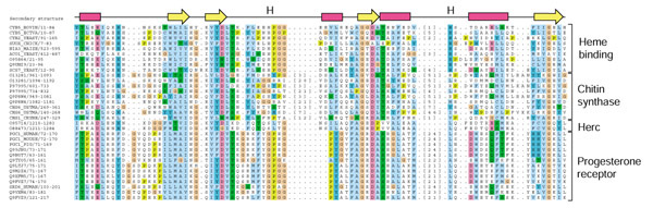
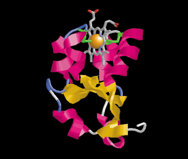
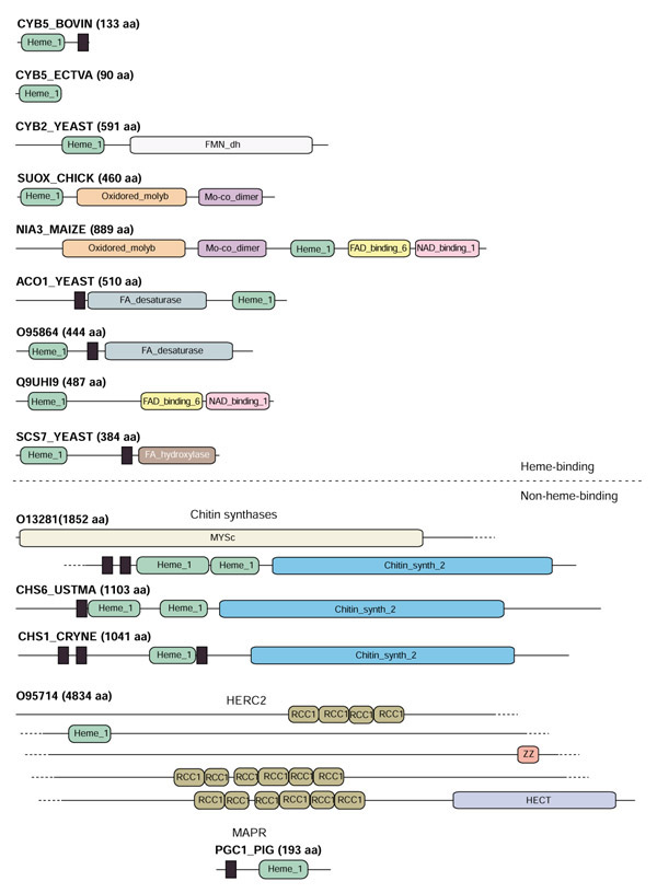
Results: We have identified MAPRs as distant homologs of cytochrome b5. We have also found regions homologous to cytochrome b5 in the mammalian HERC2 ubiquitin transferase proteins and a number of fungal chitin synthases.

Conclusions: In view of these findings, we propose that the heme-binding cytochrome b5 domain served as a template for the evolution of membrane-associated binding pockets for non-heme ligands.


Figures

Figure 1
Figure 1
Multiple sequence alignment of b5-domains. This figure has been generated using the Jalview program written by Michele Clamp. Proteins are described by their SWISS-PROT identifier or accession number/start-end, and the class they belong to is indicated on the right. Alignments are colored using the ClustalX scheme in Jalview (orange, glycine (G); light green, proline (P); blue, small and hydrophobic amino acids (A, V, L, I, M, F, W in single-letter amino acid code); dark green, hydroxyl and amine amino acids (S, T, N, Q); magenta, negatively charged amino acids (D, E); red, positively charged amino acids (R, K); dark-blue, histidine (H) and tyrosine (Y). The top line indicates secondary structural elements obtained from bovine microsomal cytochrome b5 (PDB 1cyo): magenta rectangles represent α helices, and yellow arrows represent β strands. The two positions marked 'H' indicate the conserved pair of heme-coordinating histidines in the heme-binding b5-domains. The numbers in square brackets indicate the numbers of residues not shown in this illustration.
Figure 2
Figure 2
Structure of the heme-binding domain from bovine cytochrome b5 (PDB 1cyo). The illustration has been prepared using the Rasmol program [27]. The protein is colored by secondary structure. The heme group is shown in ball-and-stick representation and the axial heme ligand histidines are shown as green side chains.
Figure 3
Figure 3
A schematic figure showing the architectures of proteins containing b5-domains. The order of the proteins in this illustration is the same as in Figure 1, except that, for proteins with a similar architecture, only one example is shown. All domain names correspond to homonymous Pfam-A families, except for the MYSc domain, which is a SMART database entry [28]. The black rectangles represent inter-domain transmembrane regions predicted by TMHMM [29]. The heme_1 domain corresponds closely to the b5-domain aligned in Figure 1. More information about each of the domains can be found in the Pfam database [18,19].

References

    1. Seyle H. Anaesthetic effects of steroid hormones. Proc Soc Biol Med. 1941;46:116–118.
    1. Falkenstein E, Norman AW, Wehling M. Mannheim classification of nongenomically initiated (rapid) steroid action(s). J Clin Endocr Metab. 2000;85:2072–2075. - PubMed
    1. Evans RM. The steroid and thyroid hormone receptor superfamily. Science. 1988;240:889–895. - PMC - PubMed
    1. Williams SP, Sigler PB. Atomic structure of progesterone complexed with its receptor. Nature. 1998;393:392–396. doi: 10.1038/30775. - DOI - PubMed
    1. Waldegger S, Beisse F, Apfel H, Breit S, Kolb HA, Haussinger D, Lang F. Electrophysiological effects of progesterone on hepato-cytes. Biochim Biophys Acta. 1995;1266:186–190. doi: 10.1016/0167-4889(94)00236-8. - DOI - PubMed

Publication types

LinkOut - more resources