

Identification of osteopontin as a prognostic plasma marker for head and neck squamous cell carcinomas
- PMID: 12538452
- PMCID: PMC12422025
Identification of osteopontin as a prognostic plasma marker for head and neck squamous cell carcinomas
Abstract
Purpose: Tumor hypoxia modifies treatment efficacy and promotes tumor progression. Here, we investigated the relationship between osteopontin (OPN), tumor pO(2), and prognosis in patients with head and neck squamous cell carcinomas (HNSCC).
Experimental design: We performed linear discriminant analysis, a machine learning algorithm, on the NCI-60 cancer cell line microarray expression database to identify a gene profile that best distinguish cell lines with high Von-Hippel Lindau (VHL) gene expression, an important regulator of hypoxia-related genes, from those with low expression. Plasma OPN levels in 15 volunteers, 31 VHL patients, and 54 HNSCC patients were quantitatively measured by ELISA. The relationships between plasma OPN levels, tumor pO(2) as measured by the Eppendorf microelectrode, freedom from relapse (FFR), and survival in HNSCC patients were evaluated.
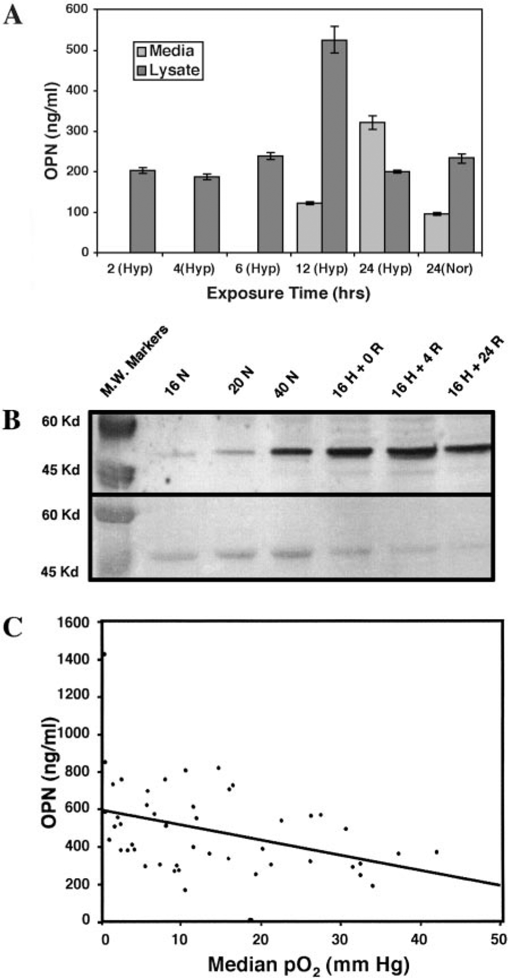
Results: Microarray analysis indicated that OPN gene expression inversely correlated with that of VHL. These findings were confirmed by Northern blot analysis. ELISA studies and Western blot in a HNSCC cell line demonstrated that hypoxia exposure resulted in increased OPN secretion. Patients with VHL syndrome had significantly higher plasma OPN levels than healthy volunteers. Plasma OPN level inversely correlated with tumor pO(2) (P = 0.003, r = -0.42). OPN levels correlated with clinical outcomes. The 1-year FFR and survival rates were 80 and 100%, respectively, for patients with OPN levels <or=450 ng/ml and 43 and 63%, respectively, for levels >450 ng/ml (P = 0.002 and 0.0005). Multivariate analysis revealed that OPN was an independent predictor for FFR and survival.
Conclusions: Plasma OPN levels appeared to correlate with tumor hypoxia in HNSCC patients and may serve as noninvasive tests to identify patients at high risk for tumor recurrence.
Figures


Comment in
-
Commentary re: Q-T Le et al, identification of osteopontin as a prognostic marker for head and neck squamous cell carcinomas. clin cancer res, 9: 31-32, 2003.Clin Cancer Res. 2003 Jan;9(1):31-2. Clin Cancer Res. 2003. PMID: 12538448 No abstract available.
References
-
- Greenlee RT, Hill-Harmon MB, Murray T, and Thun M Cancer statistics, 2001. CA - Cancer J. Clin, 51: 15–36, 2001. - PubMed
-
- Takes RP, Baatenburg de Jong RJ, Schuuring E, Hermans J, Vis AA, Litvinov SV, and van Krieken JH Markers for assessment of nodal metastasis in laryngeal carcinoma. Arch. Otolaryngol. Head Neck Surg, 123: 412–419, 1997. - PubMed
-
- Piccirillo JF, and Feinstein AR Clinical symptoms and comorbidity: significance for the prognostic classification of cancer. Cancer (Lond.), 77: 834–842, 1996. - PubMed
-
- Koch WM, Brennan JA, Zahurak M, Goodman SN, Westra WH, Schwab D, Yoo GH, Lee DJ, Forastiere AA, and Sidransky D p53 mutation and locoregional treatment failure in head and neck squamous cell carcinoma. J. Natl. Cancer Inst. (Bethesda), 88: 1580–1586, 1996. - PubMed
-
- Brennan JA, Mao L, Hruban RH, Boyle JO, Eby YJ, Koch WM, Goodman SN, and Sidransky D Molecular assessment of histopathological staging in squamous-cell carcinoma of the head and neck. N. Engl. J. Med, 332: 429–435, 1995. - PubMed
Publication types
MeSH terms
Substances
Grants and funding
LinkOut - more resources
Full Text Sources
Medical
Research Materials
Miscellaneous